

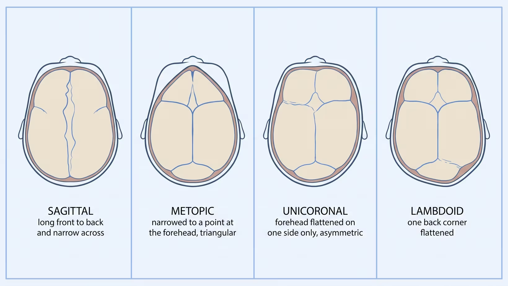
Craniosynostosis is named for the skull seam that closed too early, and the seam decides the head shape. Sagittal (top of the head) makes it long and narrow, metopic (forehead) triangular, coronal (over the top, ear to ear) lopsided, and lambdoid (back) flattens one corner. It may involve one seam or several, or be part of a genetic syndrome.
Sagittal Craniosynostosis: Why Is My Baby’s Head Long and Narrow?
Sagittal craniosynostosis is early closure of the seam that runs front to back along the top of the head, and it leaves the head long from front to back and narrow from side to side. A baby’s skull is not one solid bone. It is several plates with flexible seams between them, called sutures, and the skull grows by adding bone at those seams. Close one and the skull can no longer widen across it, so it grows the ways still open to it.
The described shape is a long, narrow, boat-shaped head with a greater front-to-back measurement, often with the forehead or the back of the head standing out. Many parents see the outline from above before anyone measures anything, and some can feel a firm ridge along the midline.
This is the most common single-seam type. In a Finnish national registry covering 877 cases, sagittal synostosis was the most common, at 3.9 per 10,000 live births, followed by metopic at 0.6, one-sided coronal at 0.4, and one-sided lambdoid at 0.1. Those are Finnish numbers, not Filipino ones, and we come back to that below. We cover the long, narrow head that sagittal synostosis makes in more detail on its own page.
Metopic Craniosynostosis: What Makes a Forehead Look Triangular?
The metopic seam runs down the middle of the forehead, from the soft spot to the top of the nose, and when it closes too early the forehead narrows to a point. Seen from above, the head has a triangular shape, and the eyes often sit closer together than usual.
Unlike the others, the metopic seam is meant to close during infancy. A ridge you can feel along the middle of the forehead is listed among the benign variations of the infant skull, meaning harmless and not cancer. A ridge on its own is not the diagnosis. What a doctor looks for alongside it is the narrowed, pointed forehead and the eye spacing.
Hindi po ibig sabihin na may sakit ang anak ninyo kapag may nakapa kayong linya sa noo. It means the head is worth looking at properly by someone who examines infant skulls often. We set the two side by side in how a harmless metopic ridge is told apart from metopic synostosis.
Coronal Craniosynostosis: What Does a Lopsided Forehead Mean?
The coronal seams run over the top of the head from ear to ear, one on each side, and when one of them closes early the forehead is flattened on that side. It is the type most likely to show in the face and not only in the skull.
One coronal seam
The forehead looks flat on the affected side and fuller on the other. The eye socket on the flat side is pulled, so the eyebrow can sit higher and the eye can look wider, and the tip of the nose can point away from that side. Parents often describe it as the two halves of the face not matching in photos.
Both coronal seams
When both coronal seams close, the skull cannot grow forward, so it grows upward and outward instead. The result is a head that is short from front to back, wide, and tall. Involvement of both sides raises the question of a syndrome more often than a single seam does, which is a question we come back to below.
For more on this type, see the lopsided forehead of coronal synostosis.
Lambdoid Craniosynostosis: Rarest Type, Mistaken for Flat Head
The lambdoid seams run across the back of the head, and early closure of one flattens that back corner. It is the rarest of the four in every series we could find, and it is the type most often confused with an ordinary positional flat head, because both flatten one side at the back.
The described findings are flattening of the back and side of the skull on the affected side, a prominent bony bump behind the ear, and an ear that sits further back and lower on that side. That is an examination finding, not a home test: the doctor looks from above and from behind and compares where the two ears sit.
Kaya po mahalaga ang pagpapatingin dito. The two are managed completely differently, and telling them apart is the point of the visit. See why a lambdoid fusion is not just a flat head.
Seek care now
When Should a Head Shape Be Seen Urgently?
Go to the nearest emergency room now for the signs in the first list below. For the signs in the second list, and for a head shape that is getting more obvious over the weeks rather than settling, have your baby seen by a pediatrician within days, not at the next scheduled well-baby check-up. Left untreated, craniosynostosis can affect development, through restricted room for the growing brain and raised pressure inside the head.
Go to the nearest emergency room now, day or night, if your baby has any of these:
- Vomiting together with unusual sleepiness or a baby who is hard to wake
- A soft spot that feels tense while your baby is calm and held upright
- Eyes that will not follow a face, or that drift
- A baby who stops doing things she was already doing
Have your baby seen by a pediatrician within days, not at the next scheduled check-up, for any of these:
- A firm ridge you can feel along a seam at the top, the side or the back of the head
- A shape that is clearer at four months than it was at one month
- A soft spot that closes very early together with a head shape that is changing
- Head circumference crossing lines on the growth chart, in either direction, over successive visits
Bring and ask
What to Bring and What to Ask at the Referral
Bring:
- The baby book with every head circumference written in it
- Photos of the head from directly above and from both sides, taken at different ages
- Any scan report, and the images or the disc, not only the printed report
- The birth history, including whether it was a long labour or a breech
Ask:
- Which seam is involved, and how sure are we
- Is this being watched or treated, and what decides that
- Does my baby need imaging, and which kind
- Is my baby still inside the window where the smaller operation is possible
- Who else is on the team, and when do we come back
How the Fused Seam Decides the Head Shape
The skull grows at its seams, so a seam that has fused stops growth across that line, and the skull compensates by growing more in the directions still open. That single rule produces all four shapes, and it is why the shape tells an experienced examiner which seam to suspect before any scan.
Read it off the map. The sagittal seam runs front to back, so losing it costs width and the head grows long. The metopic seam runs down the forehead, so losing it brings the forehead to a point. The coronal seams run across, so losing one costs forward growth on that side and losing both pushes growth upward instead. The lambdoid seams sit at the back, so losing one flattens that corner.
Multiple Suture Craniosynostosis: More Than One Seam Fused
When more than one seam is fused, the skull has fewer directions left to grow in, so pressure inside the head becomes the central concern and the surgery is usually larger. In that Finnish registry, about 83 in 100 cases involved a single seam, about 10 in 100 were craniosynostosis syndromes, and about 7 in 100 were multiple seams without a syndrome.
The pressure difference has been measured. In a series of 50 children whose pressure was measured in the operating room before their skull was opened, each additional major seam involved was linked to higher pressure, and children with more than one seam had a significantly higher median pressure than those with one. Among single-seam children in that series, metopic sat lowest and sagittal highest.
In a separate group of children examined with retinal imaging at their first vault expansion, changes suggesting raised pressure were present in 41 percent of those with one fused seam and 75 percent of those with more than one.
For a parent the meaning is simple. More seams means pressure is watched more closely and the operation planned is larger, and it is a reason to be under a craniofacial team rather than followed casually.
Syndromic vs Nonsyndromic Craniosynostosis: The Difference
Nonsyndromic means the skull is the only thing affected. Syndromic means the early fusion is one feature of a wider genetic condition that also involves the face, the hands and feet, or the airway. In the Finnish registry, about 10 in 100 cases were syndromes.
The named syndromes a craniofacial team sees include Apert, Crouzon, Pfeiffer, Muenke and Saethre-Chotzen. The familial forms are typically passed on as an autosomal dominant trait, meaning one copy of the changed gene is enough. Children with a craniofacial syndrome are far more likely to have more than one seam involved.
That is why syndromic care is team care rather than a single operation. A multidisciplinary craniofacial team with one coordinator is the recommended structure, and close monitoring is treated as mandatory because of the increased risk of raised pressure inside the head. See what syndromic craniosynostosis involves beyond the skull.
Scaphocephaly, Trigonocephaly, Brachycephaly: What They Mean
Those words describe the shape of the head, not the cause of it. That is the single most useful thing to know about them, because the same shape word can appear on a report about a fused seam and on a report about a head that was simply flattened by lying in one position.
- Scaphocephaly: long and narrow, front to back. The shape of sagittal fusion.
- Trigonocephaly: triangular, pointed at the forehead. The shape of metopic fusion.
- Brachycephaly: short front to back and wide. Seen when both coronal seams fuse, and also in positional flattening across the whole back of the head.
- Plagiocephaly: flattened on one side, an asymmetric head. Seen in coronal and lambdoid fusion, and very commonly in positional flattening.
- Synostosis: the seam itself has fused. This is the word that says the cause.
So a report saying brachycephaly has told you the shape. A report saying synostosis has told you why.
Which Type of Craniosynostosis Needs Which Surgery?
The operation is chosen by which seam fused and by how old your baby is at the time of referral, not by how severe the shape looks in a photograph. There are two broad families of operation, and age is what decides which one is even available.
The smaller operation removes the fused strip of seam through small cuts, using an endoscope (a thin tube with a camera on the end), followed by a moulding helmet that shapes the head as it grows. It is ideally done between three and four months and generally not after six months, because by then the skull is too firm for helmet moulding to work. Minimally invasive options can be offered to most babies referred under six months, which is the real reason early referral matters.
The larger operation is open cranial vault reconstruction, in which the skull plates are lifted, reshaped and refixed. It is typically done between six and 12 months, and it is the route once the endoscopic window has closed. Springs and gradual distraction of the bone are further variations used in some centres. Which part of the vault is targeted follows the seam: in lambdoid fusion it is always the back of the skull, while in sagittal fusion surgeons differ over the front and the back.
The two routes differ in more than the incision. In one centre’s series of children with Apert syndrome, the endoscopic group were operated at a median of 2.7 months against 12.8 months for open fronto-orbital advancement, with a median operating time of 65 minutes against 285, and the open group had higher rates of blood transfusion and intensive care admission and a longer stay. Head growth reached a similar level in both.
That is one syndrome at one centre, so read it as the shape of the trade-off, not a rule for every baby.
Open cranial vault reconstruction and fronto-orbital advancement are both performed in the Philippines. Published case reports from the Philippine General Hospital in Manila describe open cranial vault reconstruction for an isolated sagittal fusion, and fronto-orbital advancement for an infant with Apert syndrome. We could find no published Philippine report of the endoscopic operation, so whether it is offered near you is a question to ask the team early, while the age window is still open.
We do not print a peso figure or a PhilHealth figure here, because costs vary by centre and go stale, so ask the team what your baby’s operation would involve and what is covered.
Adults are not outside this. Fusion happens in infancy, but an adult whose craniosynostosis was never treated still has the skull it produced, and questions about pressure, headache and appearance are reasonable at any age. Those belong with a neurosurgeon, and they are a different conversation from the infant operations above.
Frequently Asked Questions
Can More Than One Type Happen in the Same Baby?
Yes. Two or more seams can fuse in the same child, and in the Finnish registry that accounted for seven percent of cases without a syndrome, while in the operating room pressure series children with a craniofacial syndrome were far more likely to have more than one seam involved. It changes the plan, because pressure inside the head is measurably higher when more than one seam is involved.
Does the Type Decide How Serious It Is?
Partly, but not on its own. Pressure measurements in one operating room series differed between single-seam types and were higher with more seams and with older age at operation, so the number of seams and the timing both matter alongside which seam it is.
Can an Ultrasound or an X-ray Show Which Type It Is?
Ultrasound of the seams can be used as a first look without radiation. In a small retrospective review of 52 scans, all seven children who had a fusion were correctly identified and no fusion was missed, although with only seven cases the certainty around that result is limited, and the choice of imaging is your team’s to make.
Will the Head Shape Come Back After Surgery?
The skull keeps growing for years after an operation, so follow-up continues rather than ending at discharge. In syndromic craniosynostosis in particular, close monitoring is treated as mandatory because of the ongoing risk of raised pressure inside the head.
How Common Is Craniosynostosis in the Philippines?
Nobody can answer that with a Philippine number. The global pooled estimate is 5.9 per 10,000 live births, but in the review that produced it, lower-middle-income countries supplied only four percent of the studies that could be pooled, so the world figure is largely a picture of high-income countries.
What to do now
What to Do Now
If you are holding a report that names a seam, take it to a pediatric neurosurgeon or a craniofacial team, and bring the images and not only the printed report. That is the visit where the type, the timing and the options get decided together.
If you have no report yet and it is the shape that worries you, book a visit with your baby’s pediatrician within days if you can feel a ridge, if the shape is getting more obvious, or if any sign in the red-flag list above is present. If none of those applies, raise it at the next well-baby check-up.
Bring photos from above and from both sides. You can also book a consultation with a pediatric neurosurgeon yourself. Ask directly whether your baby should be seen by someone who checks head shapes often, and ask early rather than at the next milestone, because the smaller operation has a window.
If your baby is vomiting and unusually sleepy or hard to wake, has a tense soft spot while calm and upright, has eyes that drift or will not follow a face, or has stopped doing things she could do, go to the nearest emergency room now. If the shape is getting worse, or another sign in the red-flag list above is present, have your baby seen by a pediatrician within days rather than at the next check-up.
Ang hugis ng ulo ay hindi ninyo kasalanan. In most babies no cause is ever found, and there is nothing a parent can point to as the reason a seam closed early. Coming in with questions is not overreacting. Nandito po kami para sagutin ang mga iyon nang maayos.
See sources
- Karsonovich T, Das JM. Craniosynostosis. StatPearls Publishing, 2025. NBK544366.
- Vuola P, Pakkasjarvi N, Ritvanen A, Heliovaara A, Tukiainen E, Gissler M. Prevalence of craniosynostosis in Finland, 1987-2010: A population-based study. Birth Defects Research, 2024. PMID 38348760 / 10.1002/bdr2.2319.
- Shlobin NA, Baticulon RE, Ortega CA, Du L, Bonfield CM, Wray A, Forrest CR, Dewan MC. Global Epidemiology of Craniosynostosis: A Systematic Review and Meta-Analysis. World Neurosurgery, 2022. PMID 35636659 / 10.1016/j.wneu.2022.05.093.
- Kalmar CL, Zapatero ZD, Kosyk MS, Carlson AR, Bartlett SP, Heuer GG, Tucker AM, Taylor JA, Lang SS, Swanson JW. Elevated intracranial pressure with craniosynostosis: a multivariate model of age, syndromic status, and number of involved cranial sutures. Journal of Neurosurgery: Pediatrics, 2021. PMID 34560658 / 10.3171/2021.6.PEDS21162.
- Swanson JW, Xu W, Ying GS, Pan W, Lang SS, Heuer GG, Bartlett SP, Taylor JA. Intracranial pressure patterns in children with craniosynostosis utilizing optical coherence tomography. Child’s Nervous System, 2019. PMID 31848721 / 10.1007/s00381-019-04448-x.
- Hersh DS, Bookland MJ, Hughes CD. Diagnosis and Management of Suture-Related Concerns of the Infant Skull. Pediatric Clinics of North America, 2021. PMID 34247705 / 10.1016/j.pcl.2021.04.002.
- Hersh DS, Hughes CD. Syndromic Craniosynostosis: Unique Management Considerations. Neurosurgery Clinics of North America, 2021. PMID 34801135 / 10.1016/j.nec.2021.09.008.
- Governale LS. Craniosynostosis. Pediatric Neurology, 2015. PMID 26371995 / 10.1016/j.pediatrneurol.2015.07.006.
- Riesel JN, Riordan CP, Hughes CD, Karsten MB, Staffa SJ, Meara JG, Proctor MR. Endoscopic strip craniectomy with orthotic helmeting for safe improvement of head growth in children with Apert syndrome. Journal of Neurosurgery: Pediatrics, 2022. PMID 35364592 / 10.3171/2022.2.PEDS21340.
- Markiewicz MR, Recker MJ, Reynolds RM. Management of Sagittal and Lambdoid Craniosynostosis: Open Cranial Vault Expansion and Remodeling. Oral and Maxillofacial Surgery Clinics of North America, 2022. PMID 35752548 / 10.1016/j.coms.2022.01.005.
- Hall KM, Besachio DA, Moore MD, Mora AJ, Carter WR. Effectiveness of screening for craniosynostosis with ultrasound: a retrospective review. Pediatric Radiology, 2017. PMID 28213626 / 10.1007/s00247-017-3793-1.
- Bernardino JD, Jose GR. Anaesthetic management of a patient with isolated non-syndromic sagittal craniosynostosis undergoing cranial vault reconstruction in the sphinx position. BMJ Case Reports, 2025. PMID 40940035 / 10.1136/bcr-2025-266735.
- Florendo GL, Jose GRB. Optimising anaesthetic management during fronto-orbital advancement in an infant with Apert syndrome. BMJ Case Reports, 2025. PMID 40953851 / 10.1136/bcr-2025-266714.