
In cranial vault remodeling surgery for craniosynostosis, a skull seam that closed too early, the surgeon lifts off the bones of the skull’s dome, reshapes them and fixes them back wider. It takes about three to five hours through a cut behind the hairline. It usually needs a blood transfusion, and your child is then watched closely in intensive care.
What Does Cranial Vault Remodeling Surgery Actually Involve?
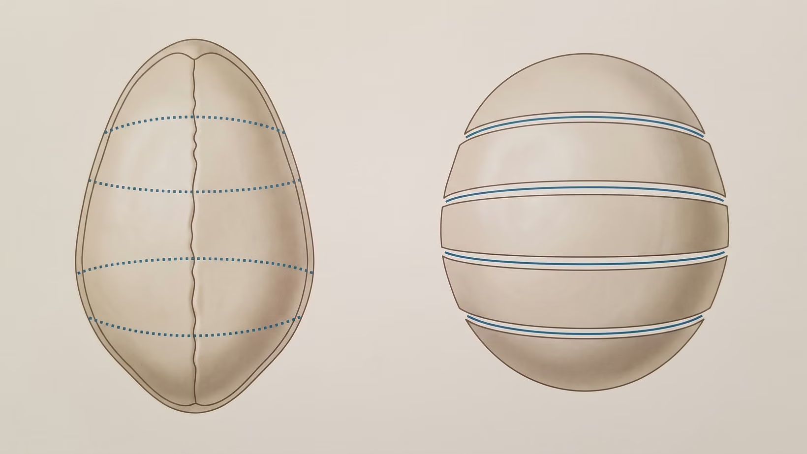
Cranial vault remodeling surgery involves lifting the affected skull bones off, cutting them into pieces, reshaping those pieces and fixing them back in a wider position. The vault is the dome of the skull above the eyebrows and ears. Remodeling means that the bone already there is rearranged into the shape the brain needs.
A suture is one of the seams between the plates of the skull. In craniosynostosis a seam closes too early. The skull cannot widen where it should, so it grows too far somewhere else instead.
Most people reading this are parents of a baby whose operation has just been booked. Some are adults who were never operated on as children. Kayo po ang magdedesisyon dito, at may karapatan kayong maintindihan ang bawat hakbang bago pumirma.
Why the Bone Is Taken Off and Reshaped, Not Just Cut
The bone comes off so that the shape is corrected in the operating room. It is not left to grow into place over the following year. Open cranial vault remodeling reshapes the existing skull bone into a wider vault. Releasing a fused seam and waiting is a different aim.
One New York team lifts a single large piece of bone off the dura, the tough membrane covering the brain. They cut it with planned saw cuts (osteotomies), shape it on a side table, and put it back as a new forehead or a new back of the head. Other teams cut and fix the bone differently. Ask your own surgeon which way they do it. I do this operation for children at Philippine Children’s Medical Center (PCMC) in Quezon City.
When the front of the skull is rebuilt, the operation can include a fronto-orbital advancement. This means the bar of bone above the eyes, called the bandeau, is taken out and moved forward too. That is why the recovery involves the eyes as well as the top of the head.
How Long the Operation Takes, and Who Is in the Room
Three to five hours is typical, and it can take longer. In a New York series of 162 children the front-of-skull operation averaged 274.9 minutes and the back-of-skull operation 216.7. In Kuala Lumpur, where children came for surgery at an older age, the operation took 6.1 hours on average.
A neurosurgeon and a craniofacial or plastic surgeon usually work together. One takes the bone off the dura and the other reshapes it.
The anesthesiologist, the doctor who puts the child to sleep, has a central role in this operation. A case report on this surgery published from the Philippines describes what the anesthesiologist manages: the airway, the position the child is placed in, breathing, keeping conditions inside the head stable, and watching blood pressure, temperature and the risk of air entering a vein.
Who Has This Operation, and at What Age
Most children have it in the first year of life, but not only then. StatPearls describes open reconstruction as typically done between six and 12 months, or when a child is referred after six months. The New York team reported first operations from three months to seven years of age.
A separate question for your own child is whether the operation is needed at all. If your baby is still under six months, ask about the smaller operation and the age by which it has to be done before this one is scheduled.
Seek care now
Which Signs After Surgery Mean Going Back to the Hospital?
These are the signs to act on in the days and weeks after you go home. While your child is still in the hospital, tell the nurse at once if you notice any of them.
If your child is unusually hard to wake, has fresh bleeding through the dressing, or vomits again and again, go to the nearest emergency room now. Tell them your child had skull surgery.
For any other sign below, call the surgical team today and bring your child in the same day. If you cannot reach them, go to the nearest emergency room and tell them your child had skull surgery. Do not wait for the follow-up appointment.
- A fever, or redness, heat or discharge at the incision. In a Vietnamese series, four of 76 children had a wound infection. Every one of them needed a return to the operating room to have the wound cleaned
- A plate becoming visible at the edge of the scar, or breaking through the skin. Three plates in a series of 134 children were exposed through the incision and had to be removed
- Fresh bleeding through the dressing, or a child who is unusually hard to wake at any time after going home. For either, go to the emergency room now, as above.
- Headache in an older child, or, at any age, repeated vomiting or the loss of a skill your child already had. For repeated vomiting, go to the emergency room now, as above. One child in the Vietnamese series needed a repeat reconstruction for raised pressure inside the head that appeared late
- A swelling under the scalp that is getting larger rather than smaller
Kung hindi po kayo sigurado, magpatingin na. Checking a wound that is fine costs you an afternoon. An infected wound that is left to wait can cost a second operation.
Where Is the Incision, and Does It Really Go From Ear to Ear?
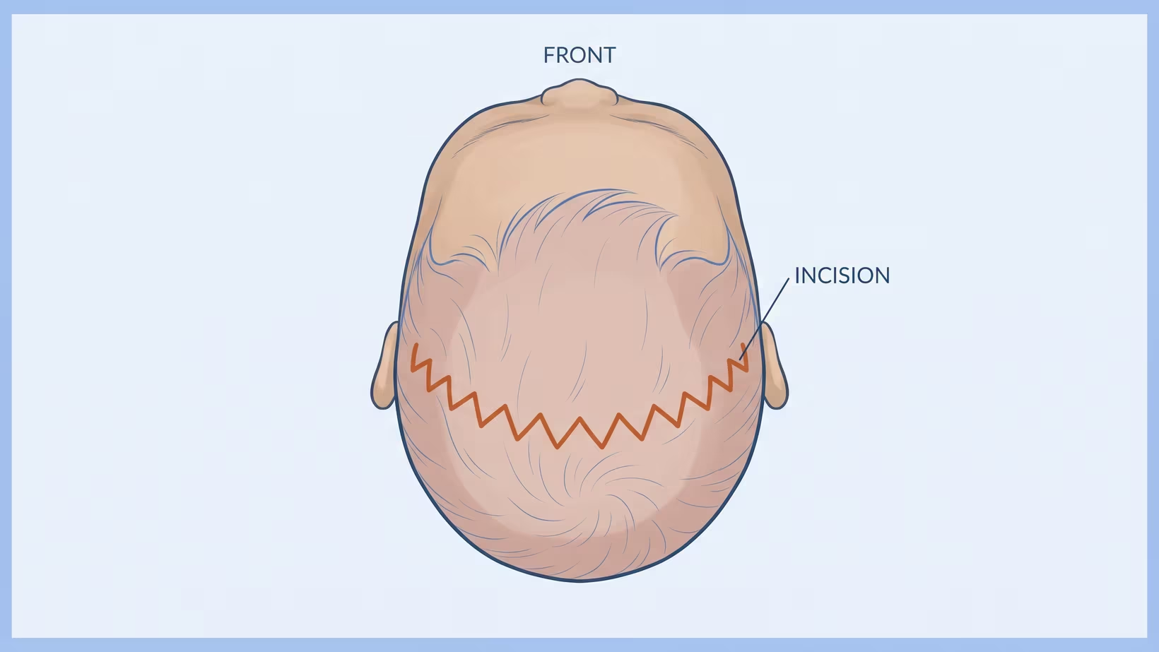
Yes, the cut for cranial vault remodeling usually goes from ear to ear. It is called a bicoronal incision. It runs across the top of the head, from just above one ear to just above the other, behind the hairline. It gives the two surgeons access to the whole dome at once.
It can be cut as a zigzag rather than a straight line. The scalp in front of and behind the cut is then lifted to expose the bone.
What the Scar Looks Like Once the Hair Grows
The scar sits behind the hairline. Even so, surgeons count a large bicoronal scar as a real difficulty for patients and families.
Thickened scars and keloids, which are raised, overgrown scars, do happen. In the New York series, 14 of the 117 children followed for a year or more had a scar revision or a keloid removed. That is about one child in eight.
Will a Blood Transfusion Be Needed?
A blood transfusion is probably needed in cranial vault remodeling. How likely it is depends greatly on where the operation is done. Pooled data from 31 North American centers (1,223 open operations) found about 91 in 100 children under two were transfused. At one high-volume American center, across 924 patients over 30 years, it was about 8 in 100.
Centers that lower the need for a transfusion describe combining several steps. These include tranexamic acid, a medicine that reduces bleeding, and recycling the child’s own blood during surgery. Ask your team about each one by name.
Older children in the published series received larger amounts of transfused blood. Part of the reason is that a bigger child has more blood to begin with. The Kuala Lumpur series also found more blood used when several seams were fused than when one was. It reported this difference as statistically significant, but the study included only 34 children.
What Are the Plates and Screws That Dissolve?
The plates and screws that dissolve are made of a material the body slowly breaks down (a resorbable polymer). They hold the reshaped bone in place, then disappear instead of staying for life. Once they are gone, the skull can keep growing. Fixed titanium hardware does not allow that.
What Happens to the Dissolving Plates Afterwards
They lose their strength and are absorbed over months while the bone heals underneath. One study used these plates in 57 children’s skull and face operations. Of the 54 upper face and midface patients, 52 healed with the bones in the intended position. The authors judged this comparable to metal plates, at a similar cost.
Failures are uncommon. Two cases have been reported in which the material did not break down as expected and had to be removed.
When Is a Lump Over the Plate a Problem?
A firm lump over a plate is common and usually goes away by itself. In a series of 134 children, about 32 in 100 had a visible lump over a plate at some point after surgery (44 children, 31.7 percent). Every one went away with watching alone. If the skin over the lump turns red or opens, or the lump keeps growing, bring your child in the same day.
Other events need attention. In that same series of 134 children, three plates came through the incision and had to be removed. These three were the series’ complications, an overall rate of about 2 in 100 children. In a separate series of 52 children, eight had a problem caused by the plate material. Seven of those eight settled without surgery, and one needed the plate removed for a skin infection.
Why Is There an ICU Stay After the Operation?
Your child stays in the ICU, the intensive care unit, because the first day is when blood pressure and body temperature can become unstable. It is also when bleeding is watched most closely. Your team will tell you why they plan it for your child.
Ang gabi sa ICU ay hindi tanda na may mali. Iyon po ang plano mula pa sa simula.
A Turkish series of 41 children after open surgery concluded that close monitoring of the heart, circulation and body chemistry in the pediatric ICU during the first 24 hours is crucial. In that series, major events occurred in about 51 in 100 children and minor ones in about 83 in 100. The paper does not define what it counted as an event. These figures are not the surgical complication rate, which is a separate and far smaller number.
How Long the Whole Hospital Stay Lasts
The whole admission lasts between two and 10 days, depending on the center. Pooled North American data put the intensive care stay at two days and the hospital stay at four. At the single high-volume center, the stays were one day and two days. The New York series averaged 5.3 days. In Kuala Lumpur, where children came to surgery older and operations took longer, the average stay was 10 days. Those days are the start of what recovery looks like from the first night onward.
Why Do the Eyes Swell Shut After Surgery?
The eyes swell shut because the operation works just above the eye sockets. The fluid it causes drains into the loose tissue around the eyes. This swelling around the eye (periorbital edema) is a known burden of the open operation, as are transfusion and fluid collecting under the scalp.
The front operation moves the bar of bone above the eyes forward, and the forehead and scalp are lifted to reach it. The eyelids have the loosest tissue on the face, so that is where the fluid gathers. The swelling does not mean the eyes themselves were operated on or harmed.
The eyes are still watched. In the Kuala Lumpur series, one of the 34 children had exposure keratitis, which is damage to the surface of the eye when the lids cannot close fully over it. You may ask how the eyes are protected during a long operation.
After you go home, bring your child in the same day if the swelling is getting worse, or if an eye looks red, waters a lot or cannot close.
What Teams Do About the Eye Swelling
Teams use the child’s position and time. Some centers also place steroids around the eyes, but this is not settled practice. Across 412 fronto-orbital advancements at two American centers, infections occurred in about 3 in 100 children given no steroid around the eyes. The figure was about 4 in 100 with an injection and about 10 in 100 with a steroid-soaked sponge. The difference may have been chance, so the question is still open.
Bring and ask
What to Bring and What to Ask Before the Operation
Bring the papers and the scans.
- The baby book or well-baby record, with every head circumference measurement in it
- Every scan already done, on a disc or a drive, not only the printed report
- The name of the seam involved, if a report already gives it
- A list of every medicine and supplement your child takes
- Your PhilHealth details, and the name of the hospital’s social service desk
Ask these questions out loud:
- How many of these operations does this team do in a year?
- What is this unit’s own transfusion rate for this operation? Is tranexamic acid or blood recycling used here?
- Will the fixation be resorbable or metal?
- How many nights in intensive care are planned, and how many in the hospital altogether?
- What will my child’s face look like the morning after, so that I am not frightened by it?
Frequently Asked Questions
Is Cranial Vault Remodeling Brain Surgery?
Cranial vault remodeling is surgery on the skull, not on the brain. It is done directly over the brain and its covering, the dura. In the Vietnamese series the most common technical event was a durotomy, a tear in that covering. It happened in about 15 in 100 cases, and none of them had an injury to the brain or the large veins.
How Big Is the Risk of Something Going Wrong?
Serious complications of cranial vault remodeling are uncommon, but they do happen. The 30-year American center reported about 7 complications in 1,000 patients. The Vietnamese series recorded wound infection in about 5 in 100 children and no deaths. The Turkish series recorded one death from bleeding among 41 children. Ask how often things go wrong at your hospital.
Will a Second Operation Be Needed Later?
A second operation is sometimes needed, and more often than the early figures suggest. In the 30-year American series, about 3 in 100 children had a second correction, at an average age of 5.5 years. By the time the skull had finished growing, that figure had risen to about 20 in 100 children.
Can This Operation Be Done on an Older Child or an Adult?
Cranial vault remodeling can be done on an older child. In the New York series, first operations were done up to seven years of age. The New York team states it can be done at any age, but their patients were children. For an adult, ask a surgeon directly.
How Much Does It Cost in the Philippines?
The cost of cranial vault remodeling in the Philippines varies by hospital and by whether the admission is service or private, so this page gives no figure. Ask the billing office for an estimate that lists the operation and the intensive care nights separately. Ask the social service desk what the operation costs here and who pays in your case.
What to do now
What to Do Now
If a date has already been given, use the weeks before it. See the surgical team at the pre-operative consult and bring the checklist above. Ask the transfusion and intensive care questions there, while the answers can still change the plan.
Ask the surgeon to describe the face you will see the morning after. Parents describe that moment as the hardest, and it is far easier when it was expected.
If no date has been given and you are still deciding whether the operation is needed, see a surgeon who treats craniosynostosis now, without waiting. The smaller endoscopic operation can only be done until about six months of age. You can book a consultation directly, or your child’s doctor can refer you.
If you would like me to see your child, there are two ways:
- A visit at my clinic at Philippine Children’s Medical Center, Room 22, Quezon Avenue corner Agham Road, Diliman, Quezon City, on Wednesday from 2 PM
- An online consult, if the trip to Quezon City is not practical for your family, booked on the consultation page
An adult can see me at the Lung Center of the Philippines, Room 1101, Quezon Avenue, Quezon City, on Tuesday and Thursday from 2 PM, or online in the same way.
Kung marami pa po kayong tanong, hindi po iyon pag-aalinlangan sa doktor. Isulat po ninyo lahat at itanong sa consult. Ang magulang na maraming tanong ay hindi po mahirap kausap. Kayo po ang katuwang namin.
See sources
- Rudy HL, Herman S, Stern CS, Staffenberg DA, Dowling K, Goodrich JT, Tepper OM. Optimizing Reconstruction in Craniosynostosis: Review of Nonsyndromic Patients Treated With a Novel Technique. Journal of Craniofacial Surgery, 2020. PMID 32569055 / 10.1097/SCS.0000000000006693.
- Tolley PD, Guyler MR, Ryan IA, Card EB, Kundishora AJ, Swanson JW. Endoscopic posterior vault distraction osteogenesis (PVDO): advantages of eliminating the bicoronal incision. Child’s Nervous System, 2026. PMID 42487007 / 10.1007/s00381-026-07391-w.
- Sakamoto Y, Arnaud E, Kamata M, Miwa T, Imai K, Sakahara D. Differences in Scalp Tension Associated With Craniosynostosis Among Patients of Different Races. Journal of Craniofacial Surgery, 2024. PMID 39356233 / 10.1097/SCS.0000000000010641.
- Wood RJ, Stewart CN, Liljeberg K, Sylvanus TS, Lim PK. Transfusion-Free Cranial Vault Remodeling: A Novel, Multifaceted Approach. Plastic and Reconstructive Surgery, 2020. PMID 31881619 / 10.1097/PRS.0000000000006323.
- Morrow BT, Ditthakasem K, Herbert M, Fearon JA. Perioperative Outcomes Following Pediatric Cranial Vault Remodeling: Are Improvements Possible? Journal of Craniofacial Surgery, 2019. PMID 31261324 / 10.1097/SCS.0000000000005675.
- Coombs DM, Ditthakasem K, Herbert M, Fearon JA. Long-Term Outcomes following Craniosynostosis Corrections: A 30-Year Retrospective. Plastic and Reconstructive Surgery, 2025. PMID 41294260 / 10.1097/PRS.0000000000012649.
- Ongun EA, Dursun O, Kazan MS, Nur B, Mihci E. Early postoperative follow-up after craniosynostosis surgery. Turkish Journal of Medical Sciences, 2018. PMID 29914256 / 10.3906/sag-1711-29.
- Can DDT, Lepard JR, Anh NM, Tuan PA, Tuan TD, Son VT, Grant JH, Johnston JM. Nonsyndromic craniosynostosis in Vietnam: initial surgical outcomes of subspecialty mentorship. Journal of Neurosurgery Pediatrics, 2021. PMID 34450594 / 10.3171/2021.5.PEDS20932.
- Koh KL, Zain A. Surgical Outcome After Less “Rigid” Fixation in Open Cranial Vault Remodeling for Craniosynostosis. Journal of Craniofacial Surgery, 2018. PMID 29438206 / 10.1097/SCS.0000000000004347.
- Witters L, Vercruysse H, De Praeter M. Total Cranial Vault Remodelling Versus Minimally Invasive Suturectomy With Postoperative Helmet Therapy in Sagittal Craniosynostosis. Journal of Craniofacial Surgery, 2024. PMID 39297663 / 10.1097/SCS.0000000000010666.
- Vega RA, Lyon C, Kierce JF, Tye GW, Ritter AM, Rhodes JL. Minimizing transfusion requirements for children undergoing craniosynostosis repair: the CHoR protocol. Journal of Neurosurgery Pediatrics, 2014. PMID 24877603 / 10.3171/2014.4.PEDS13449.
- O’Brien E, Bracken S, Yap KYB, Hunter K, Kirby F, Murray DJ. Craniosynostosis With Preoperative Iron Supplementation Exposure. Journal of Craniofacial Surgery, 2022. PMID 34411017 / 10.1097/SCS.0000000000008084.
- Sanger C, Soto A, Mussa F, Sanzo M, Sardo L, Donati PA, Di Pietro G, Spacca B, Giordano F, Genitori L. Maximizing results in craniofacial surgery with bioresorbable fixation devices. Journal of Craniofacial Surgery, 2007. PMID 17667688 / 10.1097/scs.0b013e3180a771e9.
- Wood RJ, Petronio JA, Graupman PC, Shell CD, Gear AJL. New resorbable plate and screw system in pediatric craniofacial surgery. Journal of Craniofacial Surgery, 2012. PMID 22565906 / 10.1097/SCS.0b013e31824dbed8.
- Imola MJ, Hamlar DD, Shao W, Chowdhury K, Tatum S. Resorbable plate fixation in pediatric craniofacial surgery: long-term outcome. Archives of Facial Plastic Surgery, 2001. PMID 11368657 / 10.1001/archfaci.3.2.79.
- Jerliu A, Taritsa IC, Foppiani J, Lee D, Hersh D, Lin SJ, Hughes CD. The Not Fade Away Phenomenon: 2 Cases of Delayed Nonabsorption of Biodegradable Fixation in Craniosynostosis. Plastic and Reconstructive Surgery Global Open, 2025. PMID 40040945 / 10.1097/GOX.0000000000006588.
- Massimi L, Di Rocco C. Mini-invasive surgical technique for sagittal craniosynostosis. Child’s Nervous System, 2012. PMID 22872246 / 10.1007/s00381-012-1799-4.
- Cho DY, Blum JD, Kurnik N, Swanson JW, Susarla SM, Taylor JA, Hopper RA, Bartlett S, Birgfeld CB. Eyeing Risks: A Critical Analysis of the Use of Periorbital Steroids in Fronto-orbital Advancement. Cleft Palate Craniofacial Journal, 2024. PMID 38545721 / 10.1177/10556656241241963.
- Florendo GL, Jose GRB. Optimising anaesthetic management during fronto-orbital advancement in an infant with Apert syndrome. BMJ Case Reports, 2025. PMID 40953851 / 10.1136/bcr-2025-266714.
- Bernardino JD, Jose GR. Anaesthetic management of a patient with isolated non-syndromic sagittal craniosynostosis undergoing cranial vault reconstruction in the sphinx position. BMJ Case Reports, 2025. PMID 40940035 / 10.1136/bcr-2025-266735.
- Karsonovich T, Das JM. Craniosynostosis. StatPearls Publishing, 2025. NBK544366.