
Craniosynostosis surgery is offered when a seam between the bones of the skull has fused early, and it is done to make room for the growing brain and to correct the shape that fusion has forced. It is not cosmetic surgery. Which operation your baby is offered depends mostly on age, because the smaller endoscopic route is for younger patients.
Does My Baby Really Need Surgery for Craniosynostosis?
Surgery is the usual treatment once craniosynostosis is confirmed. The reason is the risk of leaving it alone, more than the look of the head. Because of the risks linked to untreated craniosynostosis, surgery is usually done soon after diagnosis. The milder end of the range is watched instead of operated.
Alam po namin na nakakatakot ang salitang opera para sa isang sanggol. That fear is reasonable.
What the Operation Is For, and What It Is Not For
The operation exists to unlock the skull and let it grow again. The two things surgeons are trying to prevent are pressure inside the head and a shape that will not correct itself.
About 15 to 20 in 100 children with a single fused seam (15 to 20 percent) have a documented rise in pressure inside the head. About 1 in 3 children with a craniofacial syndrome do (one third). The difficulty is that the majority of patients may have neither warning signs nor symptoms for a long period of time.
When a Team Watches Instead of Operating
Not every unusual head shape leads to an operation. One British unit operated on 231 children over 20 years for a fused seam down the middle of the forehead. Surgery was offered at the moderate to severe end of the range. A ridge that can be felt, with no other feature, sat at the mild end. Kaya po may mga batang binabantayan lang muna, at hindi ibig sabihin nito na binabalewala sila.
If a team proposes watching, ask directly what they are watching for and when they will decide.
What Does Craniosynostosis Surgery Actually Do?
The operation removes or opens the fused seam, and then either lets the skull re-grow into shape or re-shapes it during the same operation. Those two halves are what separate the different techniques from each other. Removing the seam releases the restriction and gives the brain room to push the skull outward again as it grows.
Taking Out the Fused Seam
A closed seam cannot grow, so it comes out. On its own, that release is enough only while a baby’s brain is growing fast, which is why the release on its own does less the later it is done.
Reshaping the Skull
Reshaping means the bones of the vault are cut, moved and fixed in a better position during the operation itself.
Which approach a center uses depends on the seam involved, the child’s age and the team’s own experience. Ito po ang karaniwang pinag-uusapan sa unang konsulta.
Seek care now
Which Signs Mean Your Baby Should Be Seen Now?
Go to the nearest hospital emergency room now if your baby vomits repeatedly and is unusually sleepy, has a bulging soft spot, has eyes that drift or will not follow a face, has a seizure, or stops doing things they could already do. The two lists below are this clinic’s own advice to families.
Go to the nearest hospital emergency room now for any of these:
- Repeated vomiting with unusual sleepiness, or a baby who is difficult to wake
- Bumbunan, the soft spot, tense or bulging while your baby is calm and held upright
- Eyes that will not follow a face, that drift, or that suddenly turn
- A seizure
- A baby who stops doing things they were already able to do
- After an operation: fever with a red, swollen or leaking wound, or fluid collecting under the scalp
Ask for an earlier appointment, rather than waiting for the next well-baby visit, for any of these:
- A firm ridge you can feel along a seam of the skull
- A head shape that was already unusual immediately after birth
- A head shape that is more obvious at four months than it was at one month
- A head shape that has not improved at all despite changing your baby’s position
- Head circumference crossing lines on the growth chart across visits, in either direction
A normal eye examination does not rule out raised pressure inside the head, because fundoscopy and imaging are not necessarily related to what pressure recordings show. Raised pressure can cost a child their sight permanently, while the risk of it keeps rising for as long as a fused skull is left unoperated.
Endoscopic or Open Surgery: What Is the Difference?
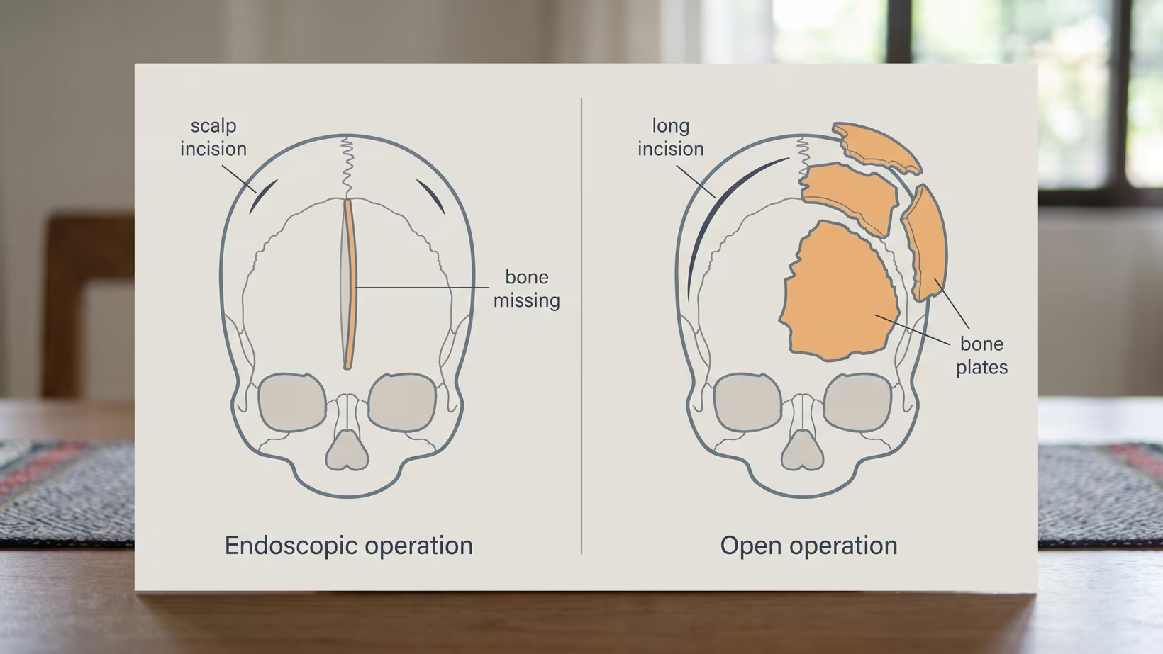
The endoscopic operation removes the fused seam through small incisions and lets the skull re-grow into shape with the help of a helmet. The open operation uses a long incision across the top of the head and re-shapes the vault in the operating room (OR) during the same sitting.
The Endoscopic Operation
It is the smaller of the two. In one single-center series of 235 consecutive babies, the median operating time was 55 minutes and the median blood loss 25 milliliters. About 96 in 100 babies (96 percent) went to an ordinary surgical ward rather than intensive care, and the median stay was one day.
A helmet is then worn for some months afterwards, and that helmet is part of the operation rather than an optional extra.
The Open Operation
Open surgery does the reshaping itself, which is why it takes longer and costs more blood. Pooling 34 studies and 11,554 patients, endoscopic surgery used about 153 milliliters less transfused blood, ran about 129 minutes shorter, and was followed by hospital stays about 2.8 days shorter.
What those same studies did not find was a difference in the shape achieved. The cephalic index, the measurement used to judge the corrected shape, came out no different between the two. On the shape they achieve, they are different operations for different ages, not a better one and a worse one.
Which One a Surgeon Offers, and Why
Age matters most, and after that the seam involved and what the center can do. Open surgery is a reasonable choice in older children. It is also the answer where an endoscope or a surgeon experienced in the technique is not available. Ask whether your center offers both.
Is There an Age Window for Craniosynostosis Surgery?
There is, and it applies to the endoscopic route rather than to surgery in general. The endoscopic operation depends on rapid brain growth to do the reshaping afterwards, so it is done in the early months. Open reconstruction works at ages the endoscopic route cannot reach.
Why the Endoscopic Window Closes
The published series operate early: across 25 studies of endoscopic repair of the seam along the top of the head, the mean age at surgery was 3.3 months.
Where the edge sits is still argued. In a review of 107 endoscopic operations on the midline seam, a quarter of babies operated after three months reached the target shape measurement. The authors still concluded that older babies may benefit.
What Is Offered After the Window
Open reconstruction is what remains. Ang mahalaga po ay hindi mawala ang pagkakataon. A worrying shape is worth showing to your pediatrician or a pediatric neurosurgeon early rather than watching for another three months.
In a national analysis of 6,010 operations, babies operated between seven and 12 months had higher odds of an in-hospital complication than those operated at zero to six months.
The Child or Adult Who Was Never Operated
A fusion that was never operated does not undo itself, and the risk of raised pressure inside the head keeps increasing for as long as there is no operation. Operating in infancy is preferable, but where that did not happen the deformity can still be corrected, as a much more complicated operation decided with the patient.
What Are the Risks, and Will My Baby Need a Blood Transfusion?
A transfusion is likely with open surgery and much less likely with the endoscopic route. In a national surgical quality database of 3,924 repairs, transfusion was the single most common recorded outcome, at about 67 in 100 patients (66.5 percent). Serious complications are uncommon and death is rare.
Blood Loss and Transfusion
Teams work at reducing blood loss. Tranexamic acid, a drug that slows the breakdown of clots, reduces both blood loss and the amount of blood transfused in this surgery, with no drug-related side effects recorded in the 258 children who received it across the pooled trials. Ask your team whether they use it.
The Other Complications
In a national study of 3,426 American repairs, about 10 in 100 babies (10 percent) had an acute complication, most often bleeding or a breathing problem. Deaths were fewer than 10 in 1,000 babies (under 1 percent).
The complications also differ by route. In a national inpatient study of 1,099 patients, surgical complications occurred in about 5 in 1,000 endoscopic cases (0.5 percent), against about 8 in 100 open cases (7.9 percent). Tears in the covering of the brain made up most of the difference, and there was no difference in deaths in hospital.
How Long Will My Baby Be in the Hospital?
Expect roughly one to two days after endoscopic surgery and three to five days after open surgery. In the national inpatient comparison the mean stay was 1.6 days for endoscopic and 3.7 days for open repairs, and a separate study of 3,426 repairs found a mean of 4.2 days.
Your own center’s answer may differ. Asking for it early helps you plan leave from work and someone to stay with your other children.
What Is Life After Craniosynostosis Surgery Like?
Most children go back to their usual daily life, with follow-up appointments and a helmet for some months if the endoscopic route was used. The outcome depends on the child. The long-term questions parents ask are about the shape, about school, and about whether a second operation is coming.
Development and School
Among 81 school-age children operated on for a fused midline seam, general intellectual ability was no different between those repaired endoscopically and those repaired openly, with adjusted mean scores of 100 and 103, and the differences from unaffected children were within the normal range.
In a five-center study of three-year-olds with a single fused seam, average scores ran three to six points below matched children, and the age at repair was not strongly related to how they scored. Kaya po nagpapa-check-up pa rin ang mga bata kahit maayos na ang hugis ng ulo.
Follow-Up, and the Chance of a Second Operation
A second operation is a real possibility rather than a likely one. In a national database of 3,924 repairs, about 2 in 100 babies (2.4 percent) had a reoperation, meaning a second operation. About 3 in 100 (3.0 percent) were readmitted to hospital. In the pooled comparison of the two routes, reoperation was less common after endoscopic surgery.
What to Send With a Craniosynostosis Referral
Send serial head circumferences with dates, photographs of the vertex, occiput and both profiles, the birth and pregnancy history, a family history of skull shape or craniofacial anomalies, and any imaging already done, on disc rather than as a report alone. Refer on the shape, not on the certainty, and refer early.
What Makes the Referral Urgent
The thresholds below are this practice’s own. Any of the emergency signs listed above means the nearest emergency room now, not a referral. Refer, rather than review in three months, for any of the earlier-appointment signs listed above, or for any of these:
- Orbital, ear or facial midline asymmetry alongside the skull shape
- Head circumference crossing centiles in either direction
- Any suspicion of a syndrome, including hand, foot, airway or midface features
What Not to Wait For
This part is for doctors referring a baby. Parents: if your pediatrician has ordered a scan, keep that appointment and ask for the referral at the same time. Do not wait on imaging to refer. Expect a clinical examination that names the seam, a decision on imaging, and a discussion of both operative routes rather than one.
Bring and ask
What to Bring and What to Ask at the Consult
Bring the baby book with every head circumference measurement, photographs of the head, any scan already done, and your PhilHealth and HMO details. Ask which seam is involved, which operation is offered, and whether your baby is still inside the endoscopic window.
Bring:
- The baby book, with every head circumference measurement in it
- Photographs of the head from directly above, from behind and from each side, taken a few weeks apart if you have them
- Any scan already done, on disc as well as on paper
- A note of anyone else in the family with an unusual head shape, or with hand, foot or hearing differences
- Your PhilHealth membership details and any HMO card
Ask:
- Which seam is involved, and is my baby still inside the endoscopic window
- Which operation are you offering, and what would change your mind
- Will a helmet be needed afterwards, and for how long
- How likely is a transfusion, and do you use tranexamic acid
- How many nights should we plan for
- What will this cost at this hospital, what does PhilHealth cover here, and who do I speak to about the balance
The cost question, including who pays for craniosynostosis surgery in the Philippines, belongs to the hospital’s own social service or billing office, not to a website.
Frequently Asked Questions
Can craniosynostosis be treated without surgery?
Craniosynostosis that needs treating is treated with an operation, because there is no medicine or helmet that opens a fused seam. The mildest end of the range is watched rather than operated. Helmets are used for flattening caused by lying position, and again as part of the plan after some endoscopic operations.
Will my baby’s head look normal afterwards?
The shape of the head usually improves substantially after craniosynostosis surgery, and the two operative routes achieve comparable measured results. The answer depends on the child. No one can promise a particular outcome for one child, which is one of the things follow-up visits exist to track.
Is one operation usually enough?
One operation for craniosynostosis is usually enough. A second operation is recorded in about 2 in 100 babies (2.4 percent) in large national datasets. In one British unit, about 6 to 7 in 100 patients (6.5 percent) had one over an average follow-up of six years.
Will the anesthesia harm my baby’s brain?
The strongest evidence on anesthesia and a baby’s brain is reassuring but does not cover this operation exactly. In a randomized trial of infants, just under one hour of general anesthesia made no difference to intelligence at five years compared with awake regional anesthesia.
Is craniosynostosis surgery done in the Philippines?
Yes, craniosynostosis surgery is done in the Philippines. Both open cranial vault reconstruction for a fused midline seam and fronto-orbital advancement in an infant with a craniofacial syndrome have been done in Manila and published in medical journals. I operate on craniosynostosis in children at the Philippine Children’s Medical Center (PCMC) in Quezon City, and my team does both the endoscopic and the open operation.
What to do now
What to Do Now
If any of the emergency signs above are present, go to the nearest hospital emergency room now and do not wait for an appointment.
You can book a consultation directly, or your pediatrician can refer you for a pediatric neurosurgery or craniofacial consultation. Bring the baby book, the photographs and any scan already done. Ask whether your baby is still inside the endoscopic window, because that is the part of this decision with a clock on it.
If you would like me to see your baby, my clinic is at PCMC, Room 22, Quezon City, on Wednesday from 2 PM. If your family lives too far for that trip, I also see families in an online consult, booked on the consultation page.
Huwag po kayong mahiyang magtanong ulit kung may hindi malinaw. Karapatan po ninyong maintindihan ang bawat hakbang bago kayo pumirma ng kahit ano.
See sources
- Governale LS. Craniosynostosis. Pediatric Neurology, 2015. PMID 26371995. DOI 10.1016/j.pediatrneurol.2015.07.006
- Tamburrini G, Caldarelli M, Massimi L, Santini P, Di Rocco C. Intracranial pressure monitoring in children with single suture and complex craniosynostosis: a review. Child’s Nervous System, 2005. PMID 15871027. DOI 10.1007/s00381-004-1117-x
- Hennedige A, Richardson D, Duncan C, et al. Part 1: surgical correction in 231 trigonocephaly patients, the Alder Hey experience. Journal of Craniofacial Surgery, 2021. PMID 33496519. DOI 10.1097/SCS.0000000000007475
- Dalle Ore CL, Dilip M, Brandel MG, et al. Endoscopic surgery for nonsyndromic craniosynostosis: a 16-year single-center experience. Journal of Neurosurgery: Pediatrics, 2018. PMID 29979128. DOI 10.3171/2018.2.PEDS17364
- Price G, Rentzeperis F, Price H, Taub P, Morgenstern P. Shaping futures: how surgical timing influences outcomes in endoscopic sagittal craniosynostosis repair. Child’s Nervous System, 2025. PMID 40369190. DOI 10.1007/s00381-025-06834-0
- De Nigris Vasconcellos F, Garcia-Torrico F, Cardoso Betta VH, et al. Evaluating surgical techniques for craniosynostosis: a systematic review and meta-analysis on endoscopic approach versus open surgery. Neurosurgery, 2025. PMID 40372012. DOI 10.1227/neu.0000000000003502
- Ortega-Ruiz OR, Torres-Martinez M, Villafranca-Cantu M, et al. Open versus endoscopic surgery with helmet molding therapy in non-syndromic patients with craniosynostosis: an updated systematic review and meta-analysis. Child’s Nervous System, 2024. PMID 39680163. DOI 10.1007/s00381-024-06692-2
- O’Brien PF, Teti SA, Dewar C, et al. Optimal timing of endoscopic sagittal suturectomy. Journal of Neurosurgery: Pediatrics, 2025. PMID 39983111. DOI 10.3171/2024.11.PEDS24272
- Abraham P, Brandel MG, Dalle Ore CL, et al. Predictors of postoperative complications of craniosynostosis repair in the National Inpatient Sample. Annals of Plastic Surgery, 2018. PMID 29489538. DOI 10.1097/SAP.0000000000001383
- Massenburg BB, Nassar AH, Hopper RA. National database reported outcomes following craniosynostosis reconstruction. Journal of Craniofacial Surgery, 2020. PMID 31764565. DOI 10.1097/SCS.0000000000006067
- Lin Y, Pan IW, Mayer RR, Lam S. Complications after craniosynostosis surgery: comparison of the 2012 Kids’ Inpatient Database and Pediatric NSQIP Database. Neurosurgical Focus, 2015. PMID 26621409. DOI 10.3171/2015.9.FOCUS15383
- Eustache G, Riffaud L. Reducing blood loss in pediatric craniosynostosis surgery by use of tranexamic acid. Neurochirurgie, 2019. PMID 31586574. DOI 10.1016/j.neuchi.2019.09.020
- Varidel A, Marucci D. Tranexamic acid in craniosynostosis surgery: a systematic review and meta-analysis. Journal of Craniofacial Surgery, 2022. PMID 34593743. DOI 10.1097/SCS.0000000000008123
- Nguyen C, Hernandez-Boussard T, Khosla RK, Curtin CM. A national study on craniosynostosis surgical repair. Cleft Palate-Craniofacial Journal, 2013. PMID 23030675. DOI 10.1597/11-324
- Kwon DY, Villavisanis DF, Choe A, et al. Complication rates and cost of endoscopic and open surgical approaches to management of craniosynostosis: a large, national, inpatient cohort evaluation. Cleft Palate-Craniofacial Journal, 2025. PMID 39980389. DOI 10.1177/10556656251320746
- Magge SN, Fotouhi AR, Allhusen V, et al. Cognitive outcomes of children with sagittal craniosynostosis treated with either endoscopic or open calvarial vault surgery. JAMA Network Open, 2024. PMID 38683606. DOI 10.1001/jamanetworkopen.2024.8762
- Starr JR, Collett BR, Gaither R, et al. Multicenter study of neurodevelopment in 3-year-old children with and without single-suture craniosynostosis. Archives of Pediatrics and Adolescent Medicine, 2012. PMID 22312170. DOI 10.1001/archpediatrics.2011.1800
- McCann ME, de Graaff JC, Dorris L, et al. Neurodevelopmental outcome at 5 years of age after general anaesthesia or awake-regional anaesthesia in infancy (GAS): an international, multicentre, randomised, controlled equivalence trial. The Lancet, 2019. PMID 30782342. DOI 10.1016/S0140-6736(18)32485-1
- Bernardino JD, Jose GR. Anaesthetic management of a patient with isolated non-syndromic sagittal craniosynostosis undergoing cranial vault reconstruction in the sphinx position. BMJ Case Reports, 2025. PMID 40940035. DOI 10.1136/bcr-2025-266735
- Florendo GL, Jose GRB. Optimising anaesthetic management during fronto-orbital advancement in an infant with Apert syndrome. BMJ Case Reports, 2025. PMID 40953851. DOI 10.1136/bcr-2025-266714
- Faasse M, Mathijssen IMJ, et al. Guideline on treatment and management of craniosynostosis: patient and family version. Journal of Craniofacial Surgery, 2022. PMID 36472893. DOI 10.1097/SCS.0000000000009143
- van Wijk RM, van Vlimmeren LA, Groothuis-Oudshoorn CGM, Van der Ploeg CPB, Ijzerman MJ, Boere-Boonekamp MM. Helmet therapy in infants with positional skull deformation: randomised controlled trial. BMJ, 2014. PMID 24784879. DOI 10.1136/bmj.g2741
- Weber J, Collmann H, Czarnetzki A, Spring A, Pusch CM. Morphometric analysis of untreated adult skulls in syndromic and nonsyndromic craniosynostosis. Neurosurgical Review, 2007. PMID 17992550. DOI 10.1007/s10143-007-0100-x
- Marchac D, Renier D, Arnaud E. Unoperated craniosynostosis patients: correction in adulthood. Plastic and Reconstructive Surgery, 2008. PMID 19050537. DOI 10.1097/PRS.0b013e31818cc388
- Becker DB, Fundakowski CE, Govier DP, Deleon VB, Marsh JL, Kane AA. Long-term osseous morphologic outcome of surgically treated unilateral coronal craniosynostosis. Plastic and Reconstructive Surgery, 2006. PMID 16525287. DOI 10.1097/01.prs.0000200613.06035.51