
An arachnoid cyst is a pocket of the clear fluid that surrounds the brain, trapped inside the thin membrane that covers it. It is not a tumor or cancer. Most are present from birth and cause no symptoms. Go to the emergency room the same day for a sudden severe headache, repeated vomiting, drowsiness, a seizure or new weakness.
What does “arachnoid cyst” mean on your scan report?
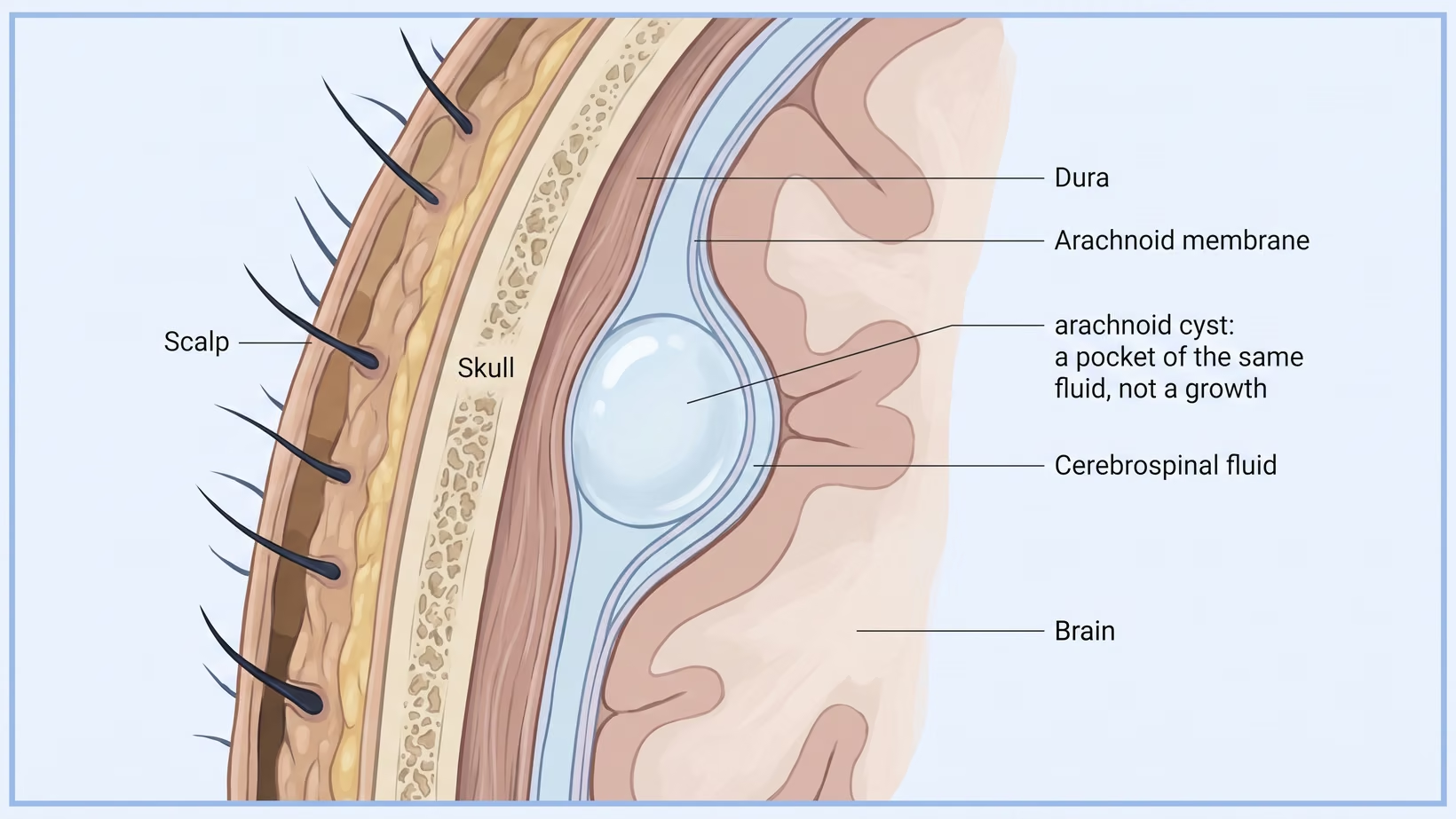
An arachnoid cyst on your report means a pocket of clear fluid between the layers of the arachnoid membrane, the thin covering over the surface of the brain. The fluid is cerebrospinal fluid (CSF), the same fluid that already cushions your brain. The pocket is filled with fluid, not with cells, so nothing inside it is dividing or spreading.
Why most arachnoid cysts are found by accident
Most arachnoid cysts are found on a scan that was ordered for something else: a headache, a head injury, dizziness, or a pre-employment medical. A finding like this is called incidental, meaning the scan found it while looking for something else. Arachnoid cysts are seen on about eight or nine out of every 1,000 brain MRI scans of people not known to have a brain problem.
Report words: extra-axial, CSF intensity, middle fossa
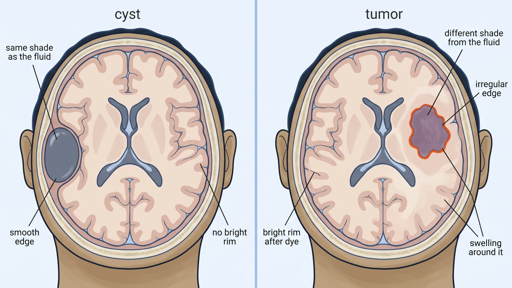
Extra-axial means the cyst sits outside the brain, beside it and not within it. CSF intensity means the fluid in the pocket looks much the same on the scan as the fluid in the normal fluid spaces, which is how the radiologist knows it is fluid and not solid tissue. Middle fossa, or middle cranial fossa, is the hollow in the base of the skull behind the temple.
Mass effect means the cyst is pressing on nearby brain. The middle fossa is one of the most common places to find an arachnoid cyst. Other fluid pockets in the head, such as pineal cysts, colloid cysts and epidermoid cysts, are different kinds of brain cyst with different names.
Is an arachnoid cyst a tumor or cancer?
No, an arachnoid cyst is not a tumor. A tumor is a mass of multiplying cells. An arachnoid cyst is a pocket of fluid, and it is not made of tumor cells.
Why some hospital websites file it under brain tumors
The word the two share is lesion, which means a spot on the scan that is not normal tissue. An unexpected finding on a brain MRI is usually not a tumor. In people with no symptoms from the brain or nerves, tumor-type findings were seen in about 7 in every 1,000 scanned, and other findings, cysts among them, in about 20.
Who tells a cyst from a tumor on a scan
The radiologist reads the scan together with your symptoms to say whether a finding is a cyst or a tumor, and the report states which. How doctors tell the two apart is set out in how a cyst differs from a tumor.
Is an arachnoid cyst dangerous?
Most arachnoid cysts are not dangerous. The large majority cause no symptoms and need no operation, because a pocket of fluid beside the brain can stay in the same place for a lifetime without doing anything. When a cyst does cause symptoms, it is because it is pressing on nearby brain or blocking the flow of brain fluid.
Symptoms an arachnoid cyst can cause
Which symptoms appear depends on where the cyst sits: headache, vomiting, a seizure, weakness or numbness on one side, double vision, unsteady walking, or in a baby a head that is growing faster than expected.
Symptoms an arachnoid cyst does not cause
A headache that is new, changing in pattern or getting worse week by week needs a doctor’s visit within the week, and any sign in the emergency list below needs the emergency room the same day. Whether your own headache is coming from the cyst is for your doctor to decide with your scan and your history.
Does the size matter, and does the location matter?
Ask the neurosurgeon to read the size together with the location and your symptoms.
When a cyst blocks brain fluid
Brain fluid is made inside the brain and drains out through narrow channels. A cyst on one of those channels can block the fluid, so the fluid spaces widen and the pressure inside the skull rises. That is hydrocephalus, which means too much fluid held inside the brain, and it is one of the main reasons an operation is considered.
When to see a doctor, and when to go to the emergency room
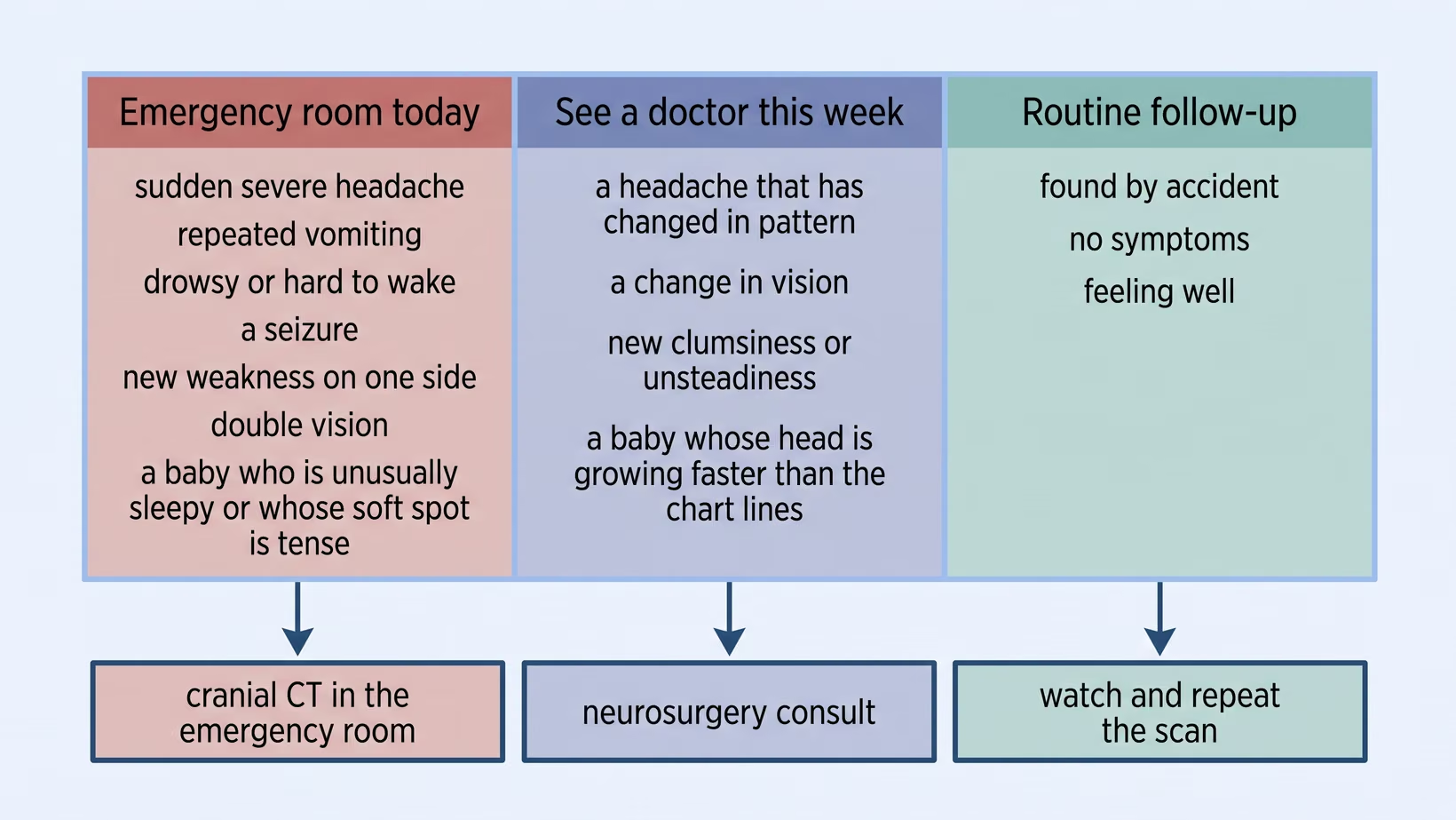
Go to the emergency room the same day for a sudden severe headache, repeated vomiting, drowsiness, a seizure, or new weakness on one side. A cyst found by accident, in someone who feels well, needs a clinic appointment and not an emergency visit.
Seek care now
Signs that need the emergency room the same day
- A sudden, severe headache that is not like your usual headache
- Vomiting again and again, especially on waking
- Drowsiness, confusion, or being hard to wake
- A seizure
- New weakness or numbness on one side of the body
- Double vision, or a new squint
- In a baby: unusually sleepy, feeding poorly, or a soft spot (fontanelle) that is tense or bulging while the baby is calm
A cyst can bleed or burst, sometimes after a head injury and sometimes without one. Watch for these signs in the days after a bump on the head.
Signs that need a check-up this week, not next year
Make an appointment within the week for a headache that has changed in pattern or is getting worse week by week, a change in vision that is not sudden, new clumsiness or unsteadiness, or a baby whose head measurement is crossing upward across the lines on the growth chart (the chart your baby’s doctor uses to plot the head measurement). A sudden loss of vision needs the emergency room the same day.
What to do now
What to bring, who to see, and what will happen at the visit
See a neurosurgeon: within the week if a symptom is changing, or at a routine appointment if you feel well. Any sign in the emergency list above means the emergency room the same day, not an appointment.
Bring the printed report, the films (the printed scan pictures), the CD or USB with the scan pictures saved on it, and a written list of your medicines. Bring one person to listen with you. The visit usually ends with a plan: watch and measure again, further tests, or an operation booked for a later date.
You may see me for this visit, or ask the doctor who ordered the scan to refer you to me.
- Adults: my clinic at the Lung Center of the Philippines, Room 1101, Quezon Avenue, Quezon City, Tuesday and Thursday from 2 PM
- Children: my clinic at the Philippine Children’s Medical Center (PCMC), Room 22, Quezon Avenue corner Agham Road, Diliman, Quezon City, Wednesday from 2 PM
- If you live too far away to come to either clinic: an online consult with me, which you book on the consultation page
Arachnoid cysts in babies, children and adults
An arachnoid cyst is usually present from birth, but it is found at every age. A baby may have one found on a scan done for head size, a child on a scan after a seizure or an injury, and an adult on a first MRI done in their forties. It is usually not something anyone caused.
Why it is usually present from birth
The arachnoid membrane forms before birth, in layers. Where the layers split instead of lying flat against each other, fluid collects in the gap. That pocket is usually already there at birth, before any fall, any fever, or anything done or not done during the pregnancy.
Found in a baby: head size, the soft spot, and feeding
A baby’s skull can still expand. A cyst that is taking up space may appear as a head growing faster than the lines on the growth chart, a soft spot (fontanelle) that feels full, or feeding that has become slow and tiring.
If the soft spot feels full or bulging while the baby is calm, or the baby is unusually sleepy or feeding poorly, go to the emergency room the same day. If the head is growing faster than the chart lines, see a doctor within the week. Keep the growth chart and bring it to the appointment.
Found in a child: seizure, head injury or school problems
Among healthy children scanned for research, about 10 in every 100 had a cyst inside the skull, counting all kinds of cyst together. So a cyst on a child’s scan may have been there all along. Whether it explains the seizure, the fall or the trouble at school is for the specialist reading that scan to say.
Found in an adult: a cyst that has been there for decades
A cyst found on a first adult scan has usually been there since birth. The neurosurgeon may ask for a repeat scan after some months to confirm that the size is not changing. For a cyst found on a pre-employment medical, the company usually needs a neurosurgeon’s note on what was found, what it means, and whether anything has to be done.
Surgery or watching: how the decision is made
Most arachnoid cysts are watched, not operated on. An operation is kept for a cyst that is causing symptoms or blocking the flow of brain fluid, so the decision rests on what the cyst is doing and not on how big it looks. Watching means a scan at a set interval, and a list of signs to report between scans.
Why watching is the usual choice
In one large population study, about 77 in every 100 people referred to a specialist for a finding on their brain MRI were watched or discharged after the first clinic visit. For cysts in the middle fossa in children, the authors of a published review concluded that surgery is needed only in patients with severe symptoms. Whether a child’s symptoms are severe enough is for the neurosurgeon who has examined the child to judge.
Fenestration and shunt, in plain words
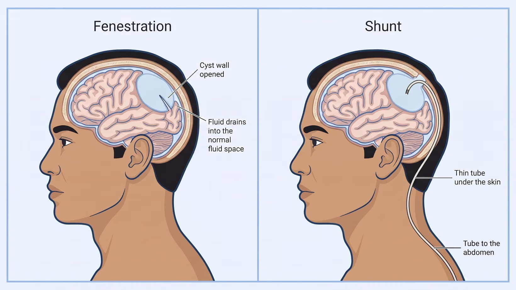
Fenestration means making an opening in the wall of the cyst, so the fluid drains into the normal fluid spaces around the brain. It is done with a microscope or with an endoscope, which is a thin tube with a camera. A shunt is a thin soft tube under the skin that carries fluid from the cyst to where the body absorbs it, usually the abdomen. Which operation fits depends on where the cyst sits and whether the fluid spaces are also enlarged.
That choice is made with a neurosurgeon who has examined you and seen your scans. I treat arachnoid cysts in children at PCMC and in adults at the Lung Center of the Philippines, and you may see me for this decision.
What can go wrong with surgery
The problems reported most often after this surgery are collections of fluid under the skull, called subdural collections and hygromas, which are pockets of fluid under the covering of the brain. After an operation, any sign in the emergency list above still needs the emergency room the same day.
Surgery carries more risk of a complication than watching does. When the neurosurgeon who has examined you advises an operation, ask about these risks at that visit rather than putting the decision off.
Living with an arachnoid cyst: follow-up scans, sports and school
A repeat scan is done at the interval the neurosurgeon sets. Between scans, and in the days after any bump on the head, a headache that keeps getting worse, repeated vomiting, drowsiness, or any other sign in the emergency list above needs the emergency room the same day.
How often the scan is repeated, and when repeat scans stop
Your neurosurgeon sets the interval, based on your age, where the cyst sits and whether you have symptoms. For cysts in the middle fossa in children, the same review concluded that after about the age of four, most children need neither surgery nor repeat scans. Keep any repeat scan your child’s neurosurgeon has already set until the neurosurgeon says it can stop.
Contact sports, boxing, diving and head protection
Whether to avoid boxing, contact sports or diving is decided case by case with the neurosurgeon. Ask the neurosurgeon before your next fight or game, not at the next routine scan. Everyone should wear a helmet when riding a motorcycle or a bicycle.
School, work, driving and the medical certificate
A cyst found by accident, with no symptoms, usually means no change to school, work or physical education unless the neurosurgeon says otherwise. Forms for driving and for some jobs usually ask about seizures and vision rather than about the cyst. Ask the neurosurgeon to write what was found, what it means and which restrictions apply.
Which cysts to refer, and what to send
Refer the same day any patient with signs of raised pressure inside the skull, new weakness, numbness or loss of vision, a first seizure, or hydrocephalus or fresh blood beside the cyst on imaging. Refer routinely when symptoms fit the cyst’s position, an infant’s head size is crossing upward on the growth chart, or a family needs the finding explained.
Refer the same day
- A reduced level of consciousness, repeated vomiting, or a headache that keeps getting worse
- New weakness on one side, a cranial nerve palsy, or double vision
- A first seizure
- Hydrocephalus on the images, or a new collection of fluid or blood (hemorrhage) next to the cyst
- An infant who is irritable or feeding poorly with a tense fontanelle
Refer routinely, and what imaging to send with the referral
Send the study itself and not only the report: the CD or USB, or the PACS link (the hospital’s online image system). Send the history too: head circumference over time in an infant, a description of the seizure, any head injury, any blood thinner (anticoagulant), and the order the symptoms started.
In a district or provincial hospital, message or call the neurosurgeon with the images and the history, rather than hold the patient for a repeat scan at the referral hospital. For a patient with any same-day sign, start the transfer to the emergency room at the same time and do not wait for a reply.
Which cysts need no referral at all
A small incidental cyst with CSF intensity, a smooth margin, no mass effect and no hydrocephalus, in a patient whose symptoms do not fit its position, needs an explanation in your clinic more than a neurosurgeon. This holds only when the MRI report calls it an arachnoid cyst, with diffusion imaging where available to rule out an epidermoid cyst. Refer if the patient is still worried after that, or if a symptom appears later that fits where the cyst sits.
Bring and ask
Cost and PhilHealth: what to ask before a scan or an operation
The cost of a scan or an operation for an arachnoid cyst differs by hospital and changes from year to year, so this page names no amounts. Ask about the PhilHealth case rate, the hospital’s estimate and assistance before admission rather than after discharge.
- Ask the attending doctor for the name of the planned procedure and its PhilHealth case rate
- Ask the billing section for the hospital’s own estimate for that procedure, including the room and the professional fees
- Ask the medical social service what assistance is available, before admission
- Bring your PhilHealth member data record and your identification, and check that your contributions are updated
- Ask what the repeat scans will cost, because follow-up is the part families forget to budget for
Frequently asked questions
Can an arachnoid cyst grow bigger?
Some arachnoid cysts do grow, so a cyst found in a baby is usually watched more closely. There is no reliable figure for how many enlarge, so the size is checked on a repeat scan.
Can an arachnoid cyst cause seizures?
An arachnoid cyst can cause a seizure, depending on where it sits, but it does not explain every seizure. Whether the cyst caused yours is for the neurologist or neurosurgeon reading the scan to decide, not the word on the report.
Did a fall, a blow to the head or hilot cause this cyst?
A fall or a blow to the head usually did not cause an arachnoid cyst. They usually form before birth, so they were usually there before any fall and before anyone touched the head. A head injury can make an existing cyst bleed, but that is different from causing it. If the head was hit hard, go to the emergency room the same day for any sign in the emergency list.
Inherited arachnoid cyst: should other children be scanned?
An arachnoid cyst found by accident, on its own, is not a reason to scan the brothers and sisters. Scanning a well child with no symptoms just to look is not advised. If a doctor finds other features that suggest a wider genetic condition, the pediatrician decides who else needs checking.
Can an arachnoid cyst go away on its own?
An arachnoid cyst sometimes shrinks on its own. This has been reported in some children whose cysts were watched rather than operated on. No figure can be given for how often, so it is not something to rely on or a reason to delay a needed operation.
See sources
- Sunny DE, et al. Prevalence of incidental intracranial findings on magnetic resonance imaging: a systematic review and meta-analysis. Acta Neurochirurgica, 2022. PMID 35525892.
- Morris Z, et al. Incidental findings on brain magnetic resonance imaging: systematic review and meta-analysis. BMJ, 2009. PMID 19687093.
- Dangouloff-Ros V, et al. Incidental Brain MRI Findings in Children: A Systematic Review and Meta-Analysis. American Journal of Neuroradiology, 2019. PMID 31624116.
- Bos D, et al. Prevalence, Clinical Management, and Natural Course of Incidental Findings on Brain MR Images: The Population-based Rotterdam Scan Study. Radiology, 2016. PMID 27337027.
- Gibson LM, et al. Potentially serious incidental findings on brain and body magnetic resonance imaging of apparently asymptomatic adults: systematic review and meta-analysis. BMJ, 2018. PMID 30467245.
- Jenkins A, et al. Comparative analysis of management techniques in the treatment of pediatric middle fossa arachnoid cysts: a systematic review and meta-analysis. Clinical Neurology and Neurosurgery, 2025. PMID 41218510.