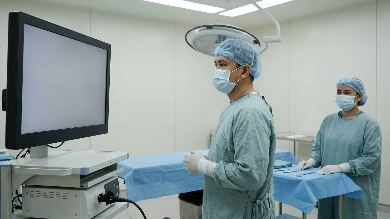

Arachnoid cyst surgery either cuts a window in the cyst wall, so the fluid drains into the normal brain fluid spaces, or places a shunt, a thin tube that drains the cyst to the abdomen. The window is cut through a camera tube or a skull opening. A worsening headache with vomiting or drowsiness needs the nearest emergency room now.
Fenestration or a shunt for an arachnoid cyst?
Fenestration means a window cut in the cyst wall, and a shunt is the other option. A review of middle fossa cysts concluded that endoscopic fenestration may be the best first operation, because it had fewer complications than the craniotomy, an open operation through the skull bone.
There are three operations for an arachnoid cyst. Two are fenestration, meaning a window cut in the cyst wall so the fluid drains into the normal brain fluid spaces. The third is a shunt that drains the cyst to the abdomen. The choice depends on where the cyst sits and whether it has already caused pressure or bleeding.
The window is cut with an endoscope, a camera tube passed through a small opening in the skull, or through a craniotomy, a flap of skull bone lifted and then put back. A review of middle fossa cysts concluded that endoscopic fenestration may be a reasonable first operation, because it had fewer complications than the craniotomy.
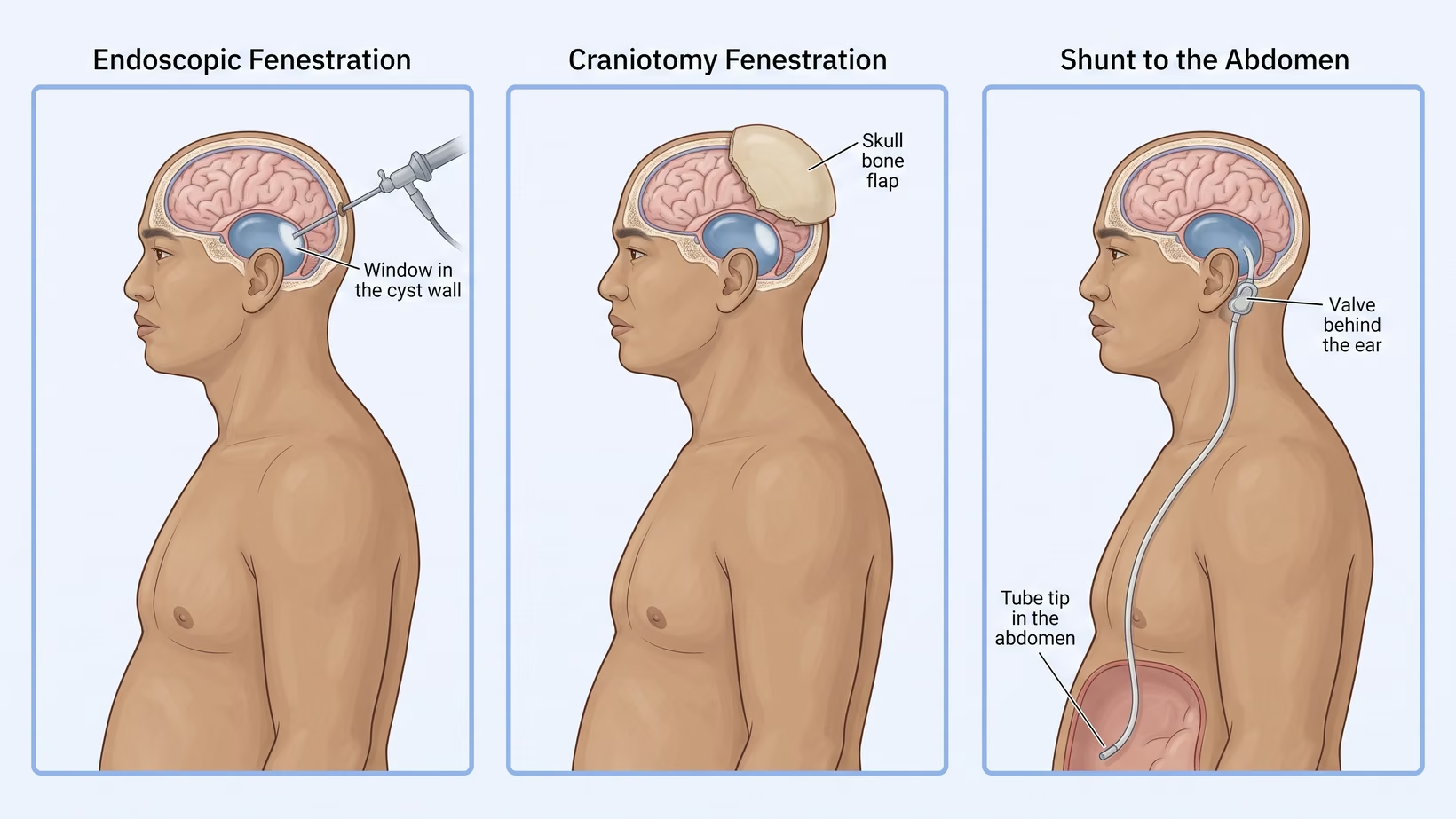
The middle fossa is the space under the temple, beside the temporal lobe. For cysts there, results added together from 18 published reports on groups of operated patients, 474 patients in all show that symptoms improved in about 90 in 100 patients after endoscopic fenestration. After fenestration through a craniotomy, symptoms improved in about 87 in 100. Only 93 shunted patients were pooled, so that group’s figure is too uncertain to compare with the other two.
The patients in these series were not alike, so the numbers do not prove that one operation is better. The figures describe patients who were already operated on. They do not tell you whether an arachnoid cyst needs surgery at all.
Seek care now
Which symptoms mean the surgery cannot wait?
Go to the nearest emergency room now for a headache that keeps getting worse with vomiting, for drowsiness or confusion, for new weakness or new double vision, or for a seizure. These are the signs of rising pressure inside the skull, and they are checked with a scan that day rather than at the next clinic appointment.
- A headache that is getting worse, with vomiting
- Hard to wake, or confused
- New weakness in an arm or a leg, or new double vision
- A seizure
- In a baby, a head growing faster than expected, or a soft spot that is tense or bulging while the baby is calm
- Any of these appearing after a bump on the head
A cyst can also tear or bleed after a fall or a bump on the head. The signs of a ruptured arachnoid cyst are the same signs, and they need to be checked in the emergency room the same way.
Bring and ask
What to bring to the neurosurgeon, and what to ask
See a neurosurgeon. With any of the warning signs above, go to the nearest emergency room now. Without them, book a consultation, or ask your doctor for a referral. Bring the scan files themselves, not only the radiologist’s report, and every earlier scan with its date, because growth is judged by comparing one scan with another.
- The CT or MRI files on a disc, a USB drive or a shared link, plus the printed report
- The dates of every previous scan, and the older scans themselves
- A written list of all medicines being taken
- The name and contact number of the doctor who referred you
Ask these four questions at that first visit. Which operation, and why that one. What happens if we wait. Who performs the operation, and in which hospital. What the follow-up looks like, and for how long.
If you would like me to see you or your child, you may come to my clinic or consult online:
- For a child: the Philippine Children’s Medical Center (PCMC), Room 22, Quezon Avenue corner Agham Road, Quezon City, on Wednesday from 2 PM
- For an adult: the Lung Center of the Philippines, Room 1101, Quezon Avenue, Quezon City, on Tuesday and Thursday from 2 PM
- If the trip is not practical: an online consult, booked on the consultation page
How is an arachnoid cyst opened with an endoscope?
The surgeon makes a small round opening in the skull bone, called a burr hole, and passes an endoscope, a thin tube with a camera and a light at its tip, down to the cyst. A window is then cut in the cyst wall, so the fluid inside drains into the normal brain fluid spaces.
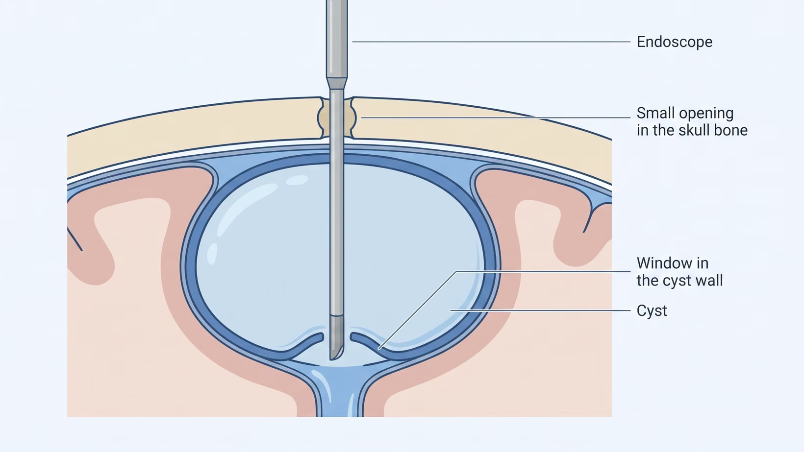
The operation does not remove the cyst. The cyst space stays where it is. What changes is that it is no longer sealed off from the rest of the fluid spaces, so pressure inside it can no longer build.
When is a craniotomy used to open an arachnoid cyst?
A craniotomy is often chosen when the cyst has torn, or when there is a collection of fluid or blood over the surface of the brain. The surgeon lifts a flap of skull bone and works under a microscope. The cyst wall is opened widely and part of it is removed, and the bone is put back.
In a review of children with middle fossa cysts, the authors judged the two operations similar overall. Symptoms from the brain improved a little more often after the craniotomy (about 99 in 100) than after the endoscope (about 91 in 100). The choice follows the situation.
What is a cyst to abdomen shunt, and when is it chosen?
A cyst to abdomen shunt, also called a cystoperitoneal shunt, is a thin soft tube that drains the cyst fluid into the abdomen, where the fluid is absorbed. Your surgeon decides from the scan whether a shunt is needed.
One end sits inside the cyst, and a valve, a small device that controls the flow of fluid, sits under the skin behind the ear. From there the tube runs under the skin of the neck and chest to the abdomen.
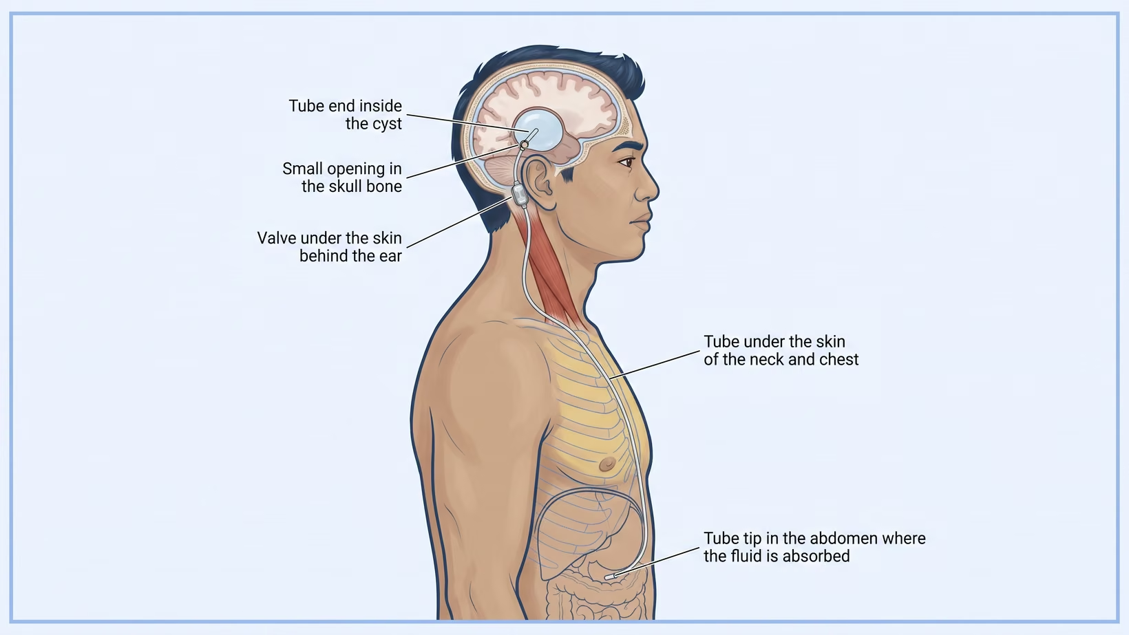
A shunt can drain the cyst, but it can block or become infected, and either one means another operation. Anyone with a shunt who has a warning sign from the box above needs the nearest emergency room now.
The clearest figures for this come from shunts placed for hydrocephalus, which is a build-up of brain fluid inside the head. In the largest randomized trial of first shunts in children (a trial in which chance decides who gets which option), about 39 in 100 shunts had failed within one year and about 53 in 100 within two years. Blockage was the usual reason and infection was next. Those were shunts in children with 1990s valves, so the figures are not the failure rate of this operation.
What are the risks of arachnoid cyst surgery?
A common problem after the cyst is opened is a collection of fluid under the covering of the brain, called a subdural hygroma. The other risks are bleeding, infection, a leak of brain fluid through the wound or the nose, and seizures. A shunt also carries the risk of problems that appear years later.
Pooled across the published series of middle fossa cysts, some complication was recorded in about 28 in 100 patients after endoscopic fenestration. The figure was about 49 in 100 after fenestration through a craniotomy, and about 20 in 100 after a cyst to abdomen shunt. Late problems, counted months and years afterwards, came to about 6 in 100 after endoscopic fenestration, about 3 in 100 after a craniotomy and about 15 in 100 after a shunt.
A complication in those series counts anything from a fluid collection that cleared on its own to a second operation. Ask your surgeon which of them could mean going back to the operating room.
In a large trial of shunts for hydrocephalus in the United Kingdom and Ireland, 32 of 533 patients with a standard shunt needed another operation for infection. With an antibiotic-coated shunt, 12 of 535 did.
How long is recovery after arachnoid cyst surgery?
Most patients stay in hospital for a few days after fenestration, and the surgeon sets the discharge day by how the patient is doing. A headache in the first days is expected and should get less each day. A headache that gets worse, or that comes with vomiting or drowsiness, is not expected: go to the nearest emergency room now, and tell the surgeon’s team on the way.
The wound is kept dry until the surgeon says otherwise. Signs of infection or of a leak of brain fluid, such as fever, a wound that turns red, swollen or starts to drain, or clear watery fluid dripping from the wound or the nose, need the surgeon’s team the same day, or the nearest emergency room if they cannot be reached. Fever with a worsening headache or drowsiness needs the nearest emergency room now.
Children usually go back to school, and adults back to desk work, once they are off strong pain medicine and sleeping normally. For most people that is a matter of weeks. The surgeon decides on contact sports, heavy lifting and driving at the follow-up visit.
What happens on the day of surgery, step by step
The anesthesiologist sees you first and tells you when to stop eating and drinking. The operation takes a few hours in most cases, and the patient wakes up in the recovery room or the intensive care unit. Most hospitals allow one bantay to stay. The ward nurse can tell you what happens before, during and after in your hospital.
Does an arachnoid cyst come back after surgery?
An arachnoid cyst can come back after surgery. A window cut in the cyst wall can scar and close again, and a shunt can block. Then a second operation is considered. For some cysts a second operation is planned from the beginning, and that is not a sign that the first one failed.
Who needs another operation depends partly on age and partly on where the cyst sits. A suprasellar cyst sits near the pituitary gland and blocks the flow of fluid. Among children operated on with an endoscope for this kind of cyst, about 29 in 100 babies under one year of age needed another operation. Among the older children, about 6 in 100 did. These figures do not apply to adults or to the middle fossa cyst.
How do doctors tell surgery worked if the cyst still shows?
The cyst usually gets smaller rather than disappearing, and a cyst still visible on the follow-up scan is the normal result. After endoscopic fenestration of middle fossa cysts, the cyst got smaller in about 76 in 100 patients, while symptoms improved in about 90 in 100. Whether the operation worked is decided on how the patient is, not on the size of the cyst.
Arachnoid cyst surgery in a child: what is different?
The operations are the same in a child; what differs is how often surgery is needed at all and how often a baby needs a second operation. Many arachnoid cysts found in children are watched and not operated on. A review of 62 published reports on middle fossa cysts in children concluded that an operation is only warranted when the child has severe symptoms. The same review found that after about the age of four most of these children need neither an operation nor repeat scans.
In a baby, head circumference measured at every visit says more than the picture does. A soft spot that is tense or bulging while the baby is calm needs the nearest emergency room now. For a suprasellar cyst, a baby under one year was more likely to need a second operation, as the figures above show. Ask your surgeon whether that applies to your child’s cyst.
I treat arachnoid cysts in children at PCMC in Quezon City.
Parents ask about anesthesia first. An international randomized trial of 722 infants compared about an hour of general anesthesia for a hernia repair with awake-regional anesthesia. The two groups scored the same on intelligence testing at five years. That was one short anesthetic for a different operation, so ask the anesthesiologist about your own child.
Is the operation different in an adult or an older person?
The operation is the same in an adult or an older person. What changes is how strictly the reason for doing it is judged. Many arachnoid cysts in adults are found on a scan done for something else. A cyst that has been there for decades without causing symptoms is not made safer by being opened.
In an older person, fluid collects more readily under the skull covering after the cyst is opened. Other illnesses and blood thinners (anticoagulants, the medicines that slow clotting) are also part of the decision, because they raise the risk of bleeding. Do not stop a blood thinner on your own; ask the surgeon and the doctor who prescribed it. A cyst that starts causing symptoms after a fall is treated with the same urgency as any bleed: go to the nearest emergency room now.
What does arachnoid cyst surgery cost in the Philippines?
No single figure covers arachnoid cyst surgery in the Philippines, and anyone who quotes one without seeing the chart is guessing. The bill is built from the doctors’ fees, the operating room, the shunt if one is used, the days in the ward or intensive care unit, the medicines and the scans.
Ask the surgeon’s secretary and the billing section for a written estimate before admission, and ask what it does not include. The medical social worker can tell you which assistance programs the hospital accepts. A private hospital bills the balance of what brain surgery costs to the family, and a government hospital starts with a social service interview.
What to send when referring for arachnoid cyst surgery
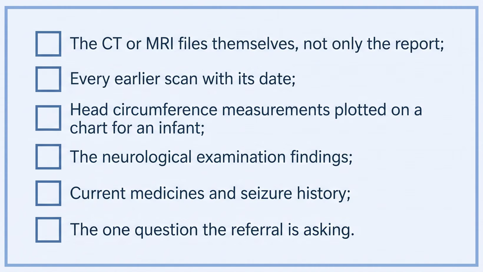
Send the images themselves, every prior study with its date, and a written neurological examination. A referral that arrives complete can be acted on at the first consult. A referral that arrives as a report alone means the patient is sent back for the files.
What to include:
- The CT or MRI on a disc, a USB drive or a shared link, not only the report. An MRI with a sequence that shows fluid, where it is available.
- Every earlier study and the date of each, so growth can be judged.
- For an infant, serial head circumference measurements plotted on a chart.
- A neurological examination with specifics: level of alertness, pupils, visual fields and eye movements, limb power, gait, and in an infant the state of the fontanelle, the soft spot on the head, and of the sutures, the seams between the skull bones.
- Seizure history, and every current medicine.
- One sentence naming the symptom that prompted the referral, and what has already been ruled out.
When it is urgent, telephone rather than write. Say the level of alertness now, what the scan shows, and how fast it has changed.
Expect back the operative plan and the reason for it, and what was discussed with the family at consent. Expect also the discharge medicines, the wound instructions, and the date of the follow-up scan.
Frequently asked questions
Can the cyst be removed completely?
No, an arachnoid cyst is not removed completely. The operation opens the cyst wall and may remove part of it, but the cyst space stays where it is. The fluid inside is no longer trapped, so pressure inside the cyst can no longer build.
Will the headaches go away?
In the published series of middle fossa cysts, symptoms as a whole improved in most operated patients. The review did not report headache on its own, and that is not a promise for any one person. Headaches have many causes, and a headache that is not coming from the cyst will not be relieved by opening it.
How long is the hospital stay?
The hospital stay after arachnoid cyst fenestration is a few days in most cases, and longer if a fluid collection or an infection appears. Your surgeon sets the discharge day by how you are doing, not by which operation was done.
Can a shunt be removed later?
A cyst to abdomen shunt can sometimes be removed later, and sometimes it cannot. It depends on whether the arachnoid cyst stays collapsed without the tube. Your surgeon judges this from the follow-up scan and from how you are doing.
Does my child need a scan every year?
Some children do not need a scan every year, but keep every follow-up scan your child’s surgeon has scheduled until the surgeon says it can stop. In the pooled series, most children past about four years of age with a middle fossa arachnoid cyst needed neither an operation nor repeat scans. Your own surgeon’s follow-up plan governs.
Which shunt valve should we ask for?
A task force review of shunt valves used for hydrocephalus in children found no design shown to be better than another, and it lists programmable and nonprogrammable valves as both options for that group. We know of no review that compares valve types for cyst shunts, so the choice belongs to the surgeon.
See sources
- Treatment of Middle Cranial Fossa Arachnoid Cysts: A Systematic Review and Meta-Analysis. World Neurosurgery, 2016. PMID 27319312. doi:10.1016/j.wneu.2016.06.046
- Comparative analysis of management techniques in the treatment of pediatric Middle fossa arachnoid cysts: A systematic review and meta-analysis. Clinical Neurology and Neurosurgery, 2025. PMID 41218510. doi:10.1016/j.clineuro.2025.109230
- Endoscopic treatment methods of suprasellar arachnoid cysts in children: A systematic review and meta-analysis. Neurosurgical Review, 2025. PMID 41286181. doi:10.1007/s10143-025-03935-x
- Drake JM, Kestle JR, et al. Randomized trial of cerebrospinal fluid shunt valve design in pediatric hydrocephalus. Neurosurgery, 1998. PMID 9696082. doi:10.1097/00006123-199808000-00068
- Mallucci CL, Jenkinson MD, et al. Antibiotic or silver versus standard ventriculoperitoneal shunts (BASICS): a multicentre, single-blinded, randomised trial and economic evaluation. The Lancet, 2019. PMID 31522843. doi:10.1016/S0140-6736(19)31603-4
- McCann ME, et al. Neurodevelopmental outcome at 5 years of age after general anaesthesia or awake-regional anaesthesia in infancy (GAS): an international, multicentre, randomised, controlled equivalence trial. The Lancet, 2019. PMID 30782342. doi:10.1016/S0140-6736(18)32485-1
- Baird LC, et al. Pediatric hydrocephalus: systematic literature review and evidence-based guidelines. Part 5: Effect of valve type on cerebrospinal fluid shunt efficacy. Journal of Neurosurgery: Pediatrics, 2014. PMID 25988781. doi:10.3171/2014.7.PEDS14325
- Rolle ML, Pascual JSG, Khu KJO, Baticulon RE, Duhaime AC, et al. Time to Care: Analysis of Time Differences and Outcomes in the Management of Pediatric Traumatic Brain Injury for a High-Income and Lower-Middle-Income Country. World Neurosurgery, 2023. PMID 37121503. doi:10.1016/j.wneu.2023.04.094