
An epidural hematoma is a bleed between the skull and the dura, the tough covering of the brain. The skull cannot expand, so the blood presses on the brain. If the person becomes harder to wake, vomits again and again, or has weakness on one side, go to the nearest emergency room now.
What is an epidural hematoma?
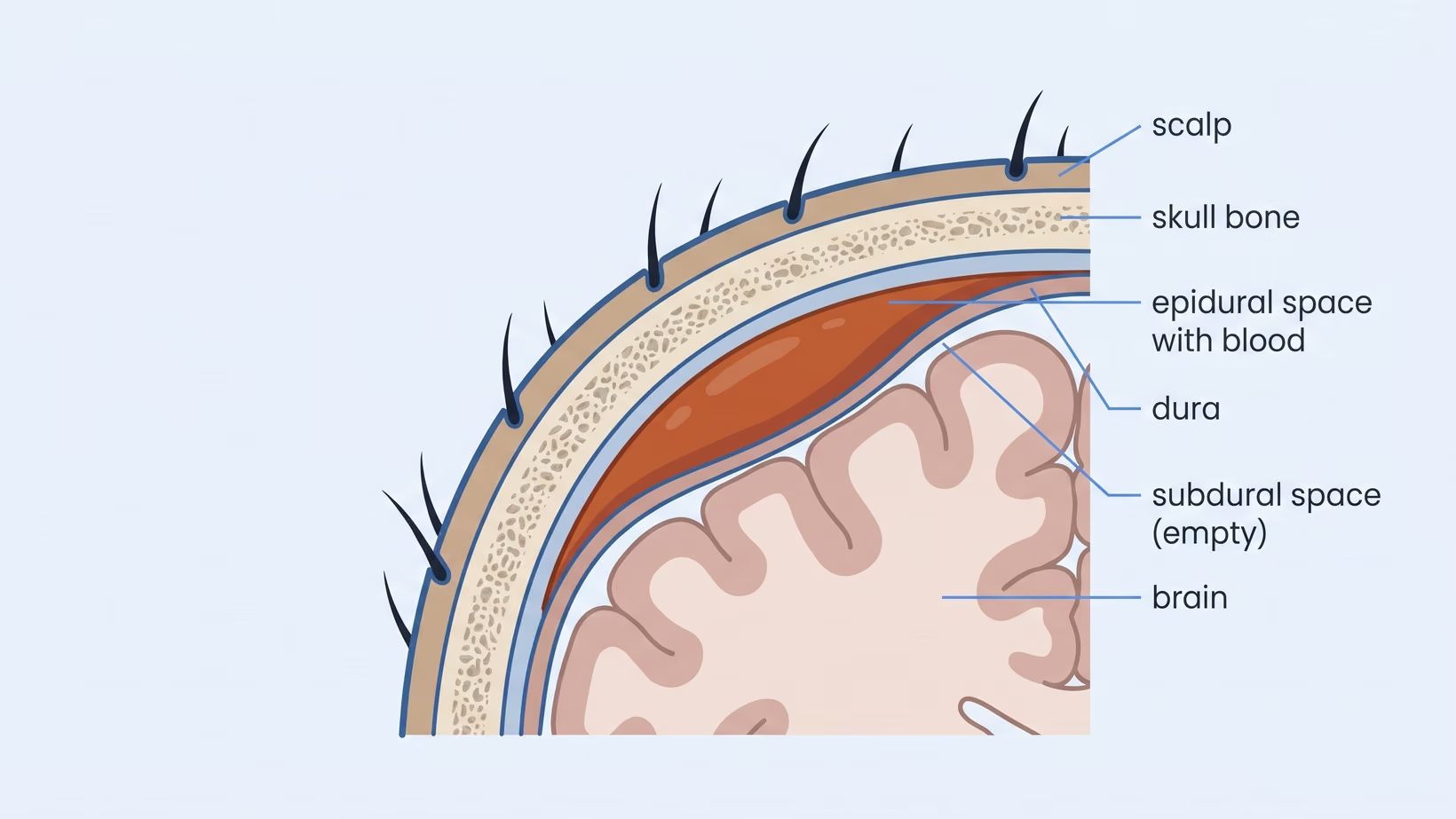
An epidural hematoma is a collection of blood between the inner surface of the skull and the dura. The dura is the tough membrane that lines the skull and wraps the brain, like a leather pouch. The skull is solid, so blood collecting there has nowhere to go. It takes space from the brain instead.
The dura is stuck to the bone, and blood has to peel it away as it collects. The brain is pushed sideways and downward. That pressure is what makes a person drowsy and then hard to wake.
Epidural hematoma symptoms: what the first hours look like
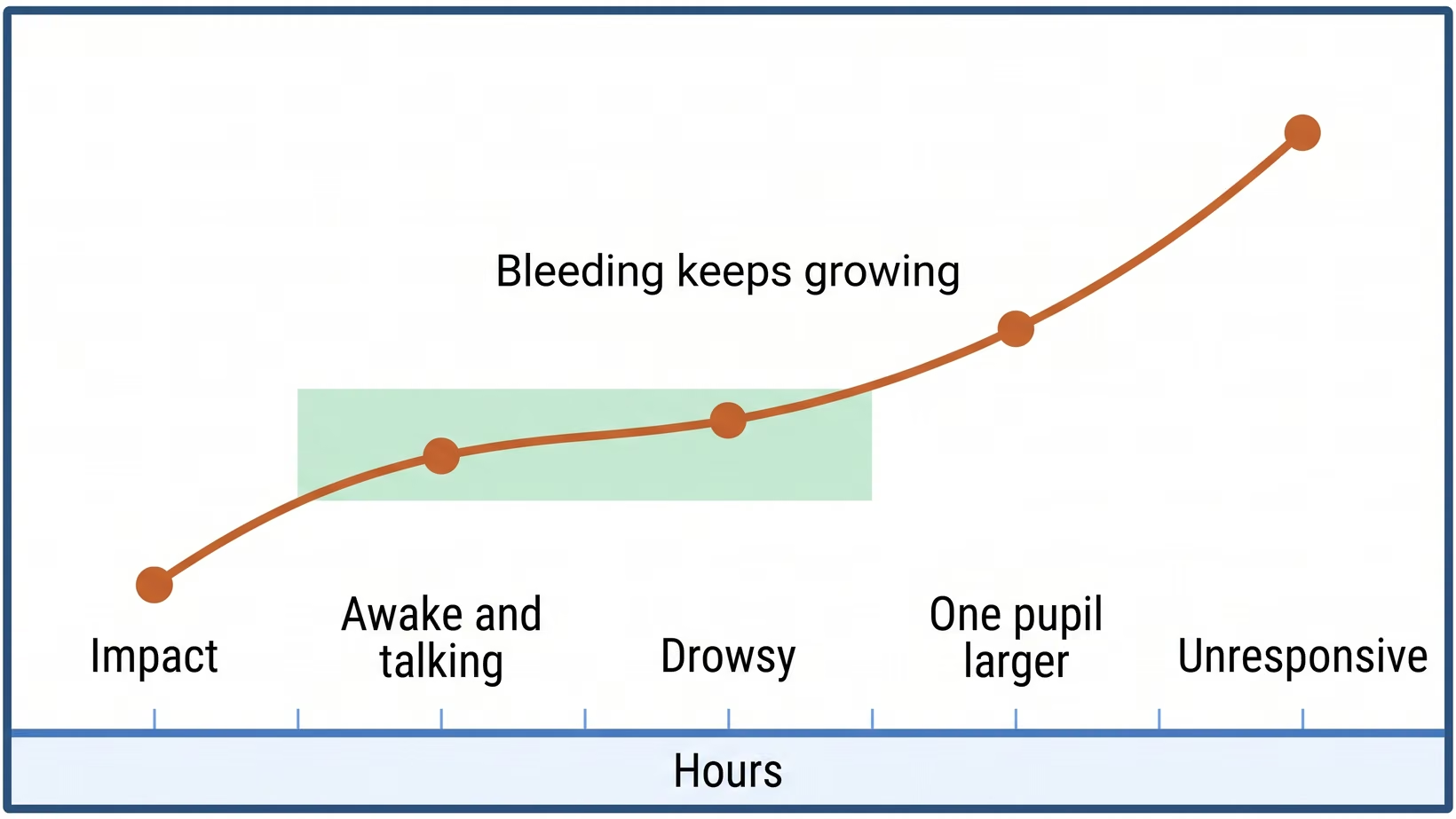
The first hours of an epidural hematoma can look mild. There is a bump to the head, sometimes a short period of being knocked out or dazed, then a headache that gets worse, vomiting, and increasing sleepiness. Later signs are one pupil larger than the other, called anisocoria, and weakness down one side of the body.
What matters is the change over time. Watch how awake the person is: slower answers, then harder to wake. At the first of these changes, go to the emergency room now; do not wait to see the next step.
Extradural hemorrhage symptoms: the same signs
Extradural hemorrhage is the same condition as an epidural hematoma, so the symptoms are the same. Hemorrhage means a bleed. If the report your hospital gives you uses that term, every warning sign on this page applies to it.
Awake and talking, then getting worse: the lucid interval
Some people are knocked out by the blow, wake up, and then talk normally for a period. Later they become drowsy and hard to wake as the clot grows. That middle period is called the lucid interval. It is the reason a person can seem fine after a head injury and then get worse.
Symptoms in a child, in an adult, and in an older person
In a child, the signs are changes in behavior: a child who will not settle, will not feed, is unusually sleepy, vomits again and again, or does not recognize you. Any of these after a head injury means going to the nearest emergency room now.
In an adult, the signs are a headache that keeps building, repeated vomiting, confusion, and weakness on one side. In an older person, sleepiness or confusion may be all the family sees. Tell the doctor every medicine the person takes, including anything that thins the blood.
Extradural hematoma and extradural hemorrhage: other names
Extradural hematoma and extradural hemorrhage are other names for an epidural hematoma: blood between the skull and the dura. Epidural means on top of the dura and extradural means outside the dura, so the two words describe one space. Some reports add an extra letter a to hematoma and hemorrhage, the British spelling of the same words.
Your CT report and your doctor may use different names, but they are not two findings. If the wording confuses you, ask the doctor to point to the collection on the film and name it once, in the words you will repeat at the next hospital.
When to go to the emergency room, and when to refer immediately
Go to the nearest emergency room now if a person who has hit their head becomes harder to wake, vomits again and again, or develops weakness on one side. If a hospital with a CT scanner is just as close, go there. Do not wait for morning and do not wait to see whether it settles. An epidural hematoma can grow while the person still looks reasonable.
Seek care now
Red flags after a head injury: go now, not in the morning
- Harder to wake than they were an hour ago, or will not wake fully
- Repeated vomiting after the injury
- A headache that keeps getting worse
- One pupil larger than the other
- Weakness, numbness or clumsiness down one side
- A seizure, or any abnormal jerking
- Confusion, agitation, or speech that makes no sense
- Clear fluid or blood coming from the nose or the ear
- In a baby or small child: will not feed, will not settle, unusually sleepy, or a bulging soft spot
- Any head injury in a person taking a blood thinner, which is a medicine that slows clotting
Do not take the person for hilot first. Go to the nearest emergency room, or to a hospital with a CT scanner if one is just as close. Time spent anywhere else is time lost.
For referring physicians: who needs a scan, and how fast
In adults who are awake after a head injury, the Canadian CT Head Rule names five high-risk findings: not reaching full alertness within two hours, a suspected open skull fracture, any sign of a fracture at the base of the skull, vomiting more than twice, and age over 65.
Full alertness here means a Glasgow Coma Scale score of 15, the score that rates how awake a person is. In the study that produced the rule, those high-risk findings identified everyone who went on to need an operation. The rule was derived in adults who arrived awake, so it does not apply to children and it does not rule an epidural hematoma in or out.
Bring and ask
What to send with the patient, and who to call first
If you are the doctor sending the patient, call first and find out which hospital near you has both a CT scanner and a neurosurgeon on duty. If you are family and the person is getting worse, do not stop to call: go to the nearest emergency room now. There are about 174 neurosurgeons in the Philippines, roughly one for every 600,000 people, and 35 provinces have none at all. The nearest hospital is not always the one that can operate.
Send with the patient:
- The scan images on a disc or a USB, and the printed report, not the report alone
- The time of the injury, written down, and the time the sleepiness started
- The Glasgow Coma Scale score on arrival and every change since
- A written list of medicines, above all anything that thins the blood
- The name and number of the doctor who is sending them
- A relative who can give consent for an operation
For the check after the hospital stay, your doctor can refer you to a neurosurgeon, or you can book a consultation yourself and bring the same papers to that first neurosurgery appointment.
You may also see me for that check. I see adults at the Lung Center of the Philippines on Tuesday and Thursday from 2 PM, and children at the Philippine Children’s Medical Center (PCMC) on Wednesday from 2 PM. If the trip is too far for you, book an online consult on the consultation page.
What an epidural hematoma looks like on a CT scan
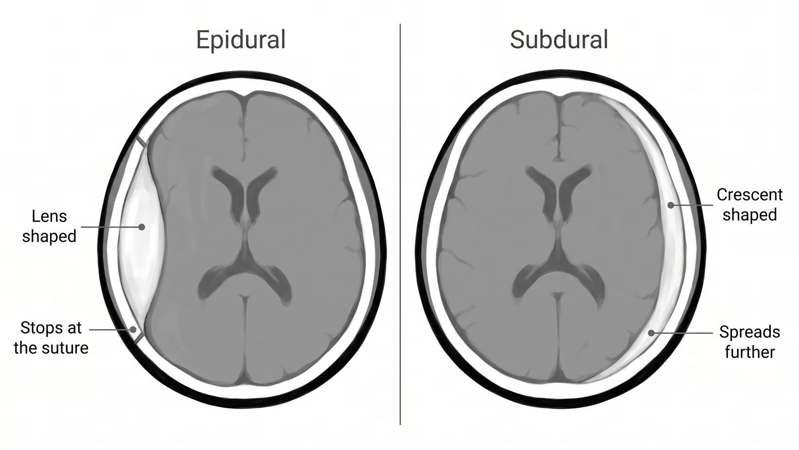
On a CT scan an epidural hematoma is a bright collection shaped like a lens: wide in the middle and narrow at both ends. It sits tight against the inside of the skull. The report calls that shape biconvex, meaning curved outward on both sides.
Why it is lens shaped and stops at the suture lines
The dura is attached to the inside of the skull, most tightly along the sutures. The sutures are the seams where the skull bones meet. Blood peels the dura off the bone between the seams, and the seams hold it back. That is what gives the collection its lens shape and its edges.
Epidural or subdural: how the shape tells them apart
A subdural hematoma sits one layer deeper, under the dura, where no seam stops it. It spreads as a thin crescent that follows the curve of the skull for a longer distance. That is why a crescent shaped bleed on a CT scan is read as a subdural, while a lens held in by the seams is read as an epidural.
Why it bleeds so fast: artery and temporal bone fracture
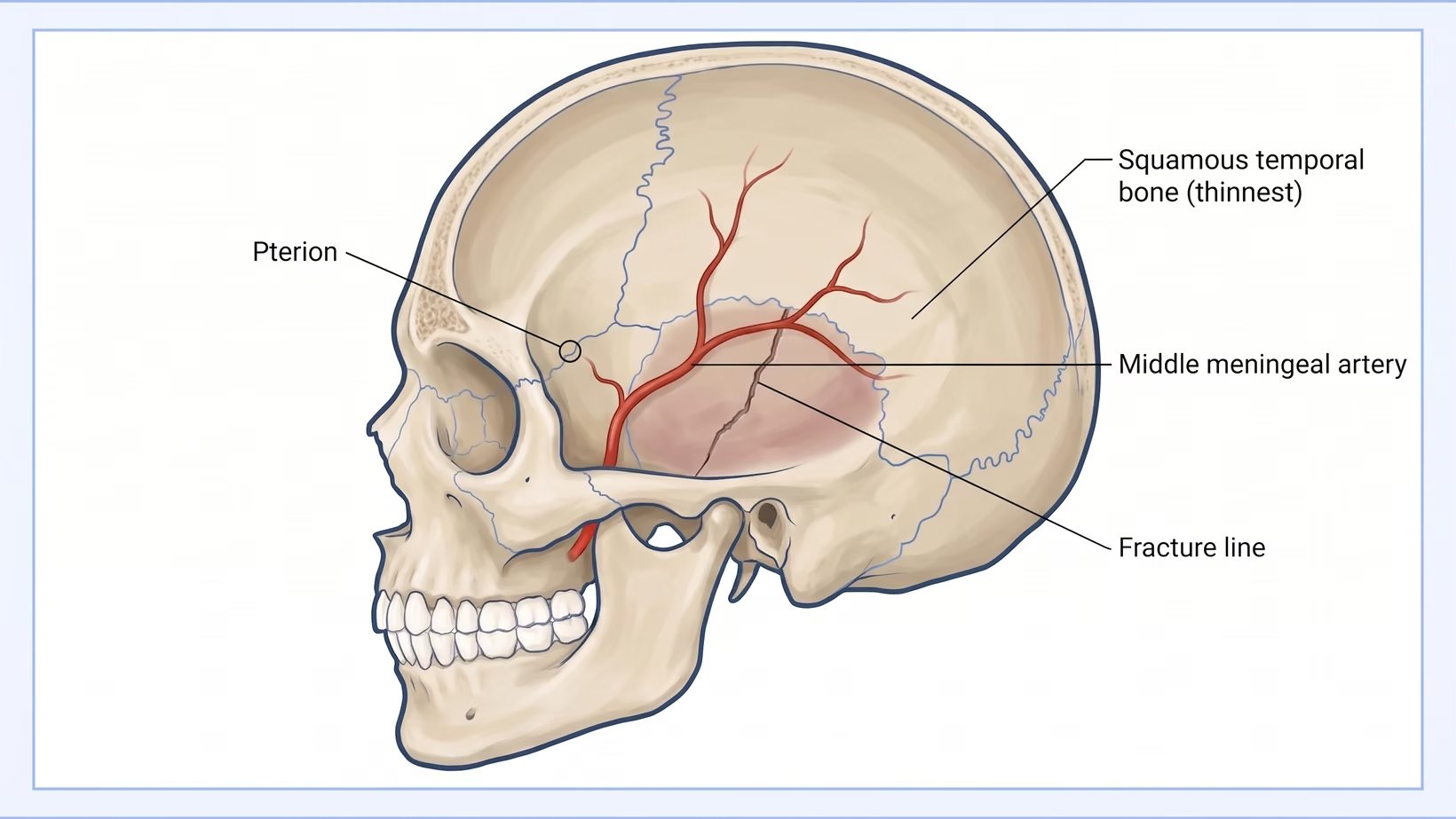
The middle meningeal artery is an artery that runs in a shallow groove on the inner surface of the skull above the ear.
The thinnest part of the skull, and the artery running under it
The squamous temporal bone is the flat part of the skull just above and in front of the ear, and it is the thinnest part of the skull. A blow to the side of the head lands on that thin bone. The artery runs underneath it, which is why a crack in that one place matters more than a crack elsewhere.
Epidural hematoma in children: what is different
Children get epidural hematomas, and it is the same emergency. In one Philippine trauma center’s series of children admitted with a head injury, an epidural hematoma was the most common abnormal finding on the scan. The most common operation was a craniotomy to remove an epidural hematoma, which means opening a window of skull bone over the clot.
Most of the children in that series had mild injuries, and most went home without complications. That is one hospital’s experience with children who were admitted, not every child who hits their head. It does not mean a child with any red flag can wait.
Why a child can stay alert, and why that is not reassurance
A child can be awake, talking and even playing while a collection of blood is already growing inside the skull.
Compare the child with how they were an hour ago, not with how a sick child looks. Any of the changes in the red flags, or a limb they have stopped using, is a reason to go now.
Which children need a CT scan after a head injury
Many children who hit their head do not need a CT scan, and the reason to avoid one is the radiation. A child with any red flag should still be brought in now. A large study found a group of children at very low risk, and the doctor who examines the child uses its signs to decide which children need a CT scan and which can be watched.
Epidural hematoma after a motorcycle crash
A motorcycle crash can put the side of the head onto the road or onto another vehicle, and the side is where the skull is thinnest. Road crashes were the main cause of injury in the same Philippine series of children. The injury happens away from a hospital, and the time that matters starts at the crash.
Why riders arrive with this bleed, and what the helmet changes
A helmet lowers the risk of dying and the risk of head injury among riders who crash. That evidence comes from observational studies, not trials, and it covers head injury as a whole, not this bleed in particular. A rider who was wearing a helmet and is now drowsy still needs the scan.
The first hour in a Philippine emergency room
The first hour in the emergency room goes to breathing, blood pressure and conscious level, then to a CT scan of the head if the hospital has one. If that hospital has no scanner or no neurosurgeon on duty, the first hour is also spent arranging a transfer.
Epidural hematoma treatment: what the hospital does
The treatment for an epidural hematoma that is pressing on the brain is an operation to remove it. The surgeon opens the scalp, lifts a window of skull bone over the collection, clears the blood, and puts the bone back. Stopping the bleeding point is what keeps the collection from coming back. This operation is called a craniotomy.
The guideline does not name one preferred method, but it states that a craniotomy clears the clot more completely than the alternatives, and the surgeon chooses the approach for each patient.
Intensive care afterwards, and what is being watched
After the operation the person is watched closely, usually in intensive care, and a repeat scan is done whenever anything changes. The team checks how awake the person is, the pupils, the limbs, the breathing and the blood pressure.
Steroids are not given for the swelling of a head injury. The large trial that tested them found more deaths in the group that received them.
What the operation and the hospital stay will cost
No one can give you a reliable figure before the patient is seen. Ask the billing section for an estimate in writing once the patient is admitted, because what the operation costs depends on the hospital, the days in intensive care and what else was injured. Ask what a deposit before surgery covers and what it does not.
Surgical thresholds: when an epidural hematoma needs surgery
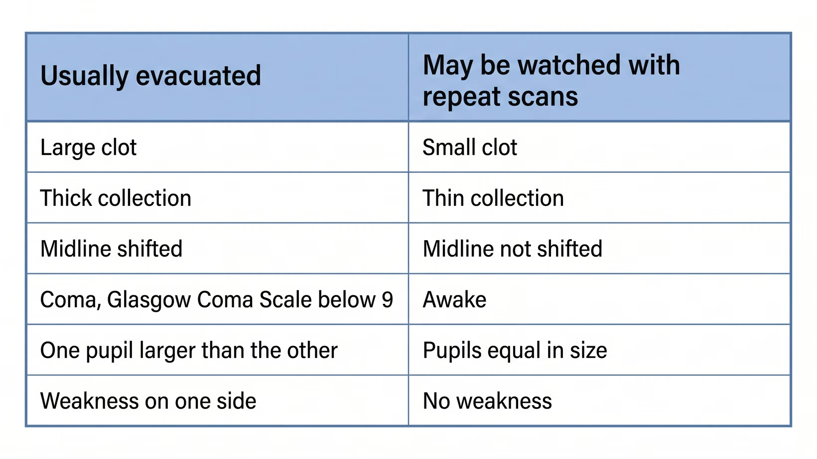
The guideline recommendation is that an epidural hematoma larger than 30 cubic centimeters is removed by an operation whatever the person’s conscious level. Thirty cubic centimeters is about two tablespoons of blood. A surgeon weighs these numbers together with the whole patient. They are not a formula, and neither side of the line is safe.
The size, thickness and midline shift that decide it
A smaller one can be watched in a neurosurgical center, with repeat scans, if it is less than 15 mm thick, pushes the midline less than 5 mm, and is in a person with a Glasgow Coma Scale score above 8 and no focal deficit, meaning no weakness or other sign pointing to one part of the brain.
Thickness is the depth of the collection at its deepest point, and 15 mm is one and a half centimeters. Midline shift is how far the middle of the brain has been pushed across, measured in millimeters. A Glasgow Coma Scale score runs from 3 to 15, and a score above 8 means the person is not in a coma.
When a small epidural hematoma can be watched instead
Watching is a hospital activity. The guideline allows it only with repeat CT scans and close neurological observation, meaning regular checks of how awake the person is, the pupils and the limbs, in a neurosurgical center. A small collection is never a reason to go home and watch. The point of watching is that an operating room is minutes away if the person changes.
Why the time from deterioration to the operating room matters
A person with an acute epidural hematoma, meaning a new one, who is in a coma, with a Glasgow Coma Scale score below 9, and who has one pupil larger than the other should have the clot removed as soon as possible. The guideline is at its strongest on this point.
No number of minutes is given. The operation comes first and the rest is arranged around it.
Frequently asked questions
Can an epidural hematoma heal on its own?
A small epidural hematoma can be watched with repeat scans and close checks in a neurosurgical center, and some do not need an operation. No medicine dissolves a clot that is already pressing on the brain. Tranexamic acid is a medicine the hospital team may give early after a head injury to help limit bleeding. It is not something to give at home, and it does not replace removing a clot.
How long is the operation and the hospital stay?
The length of an epidural hematoma operation and of the hospital stay has no single answer. Both depend on the size of the clot, the state of the brain underneath it, and what else was injured. Ask the surgeon for the expected range for this patient.
Will there be lasting damage after an epidural hematoma?
Whether an epidural hematoma leaves lasting damage depends on the whole injury and not on the clot alone. How awake the person was before the operation, what else the brain took, and what the repeat scans show all go into it. Ask the team about what the first year of recovery holds.
Is an epidural hematoma the same as a stroke?
An epidural hematoma is not a stroke. A stroke is a blocked or burst blood vessel inside the brain itself. An epidural hematoma is blood collecting outside the brain, between the skull and the dura, and it follows an injury.
Can an epidural hematoma come back after surgery?
The team watches for an epidural hematoma coming back after surgery, which is why the checks continue through the night. Any change in how awake the person is, a new weakness, or a pupil that becomes larger is a reason to repeat the scan rather than wait until morning. If you are at the bedside and notice any of these, tell the nurse at once.
Does PhilHealth cover an epidural hematoma operation?
PhilHealth benefits for an epidural hematoma operation depend on the hospital and on the case, and the amounts change, so any figure printed here would mislead you. Ask the PhilHealth desk which benefit applies to this admission, whether the hospital is a PhilHealth facility, and whether the surgeon is accredited.
See sources
- Bullock MR, Chesnut R, Ghajar J, Gordon D, Hartl R, Newell DW, Servadei F, Walters BC, Wilberger JE. Surgical management of acute epidural hematomas. Neurosurgery (Surgical Management of Traumatic Brain Injury guideline series), 2006. PMID 16710967.
- Stiell IG, Wells GA, Vandemheen K, et al. The Canadian CT Head Rule for patients with minor head injury. Lancet, 2001. doi:10.1016/s0140-6736(00)04561-x.
- Kuppermann N, Holmes JF, Dayan PS, et al (PECARN). Identification of children at very low risk of clinically-important brain injuries after head trauma: a prospective cohort study. Lancet, 2009. doi:10.1016/S0140-6736(09)61558-0.
- Liu BC, Ivers R, Norton R, Boufous S, Blows S, Lo SK. Helmets for preventing injury in motorcycle riders. Cochrane Database of Systematic Reviews, 2008. doi:10.1002/14651858.CD004333.pub3.
- Baticulon RE, Lucena LLN, Gimenez MLA, Sabalza MN, Soriano JA. The Neurosurgical Workforce of the Philippines. Neurosurgery, 2023. doi:10.1227/neu.0000000000002630.
- Bayhon MV, Sih IMY. Clinical Characteristics and Outcomes of Pediatric Traumatic Brain Injury Patients in a Tertiary Regional Trauma Center in the Philippines. Neurotrauma Reports, 2025. doi:10.1089/neur.2025.0002.
- CRASH-3 trial collaborators. Effects of tranexamic acid on death, disability, vascular occlusive events and other morbidities in patients with acute traumatic brain injury (CRASH-3): a randomised, placebo-controlled trial. Lancet, 2019. doi:10.1016/S0140-6736(19)32233-0.
- Roberts I, Yates D, Sandercock P, et al (MRC CRASH trial collaborators). Effect of intravenous corticosteroids on death within 14 days in 10008 adults with clinically significant head injury (MRC CRASH trial): randomised placebo-controlled trial. Lancet, 2004. doi:10.1016/S0140-6736(04)17188-2.