

Bleeding in the brain, also called a hemorrhagic stroke, is a stroke caused by blood leaking from a vessel into the brain or the space around it. It is an emergency, so take the person to the nearest emergency room now. A brain scan is needed to tell a bleed from a clot, because each is treated differently.
What is a hemorrhagic stroke?
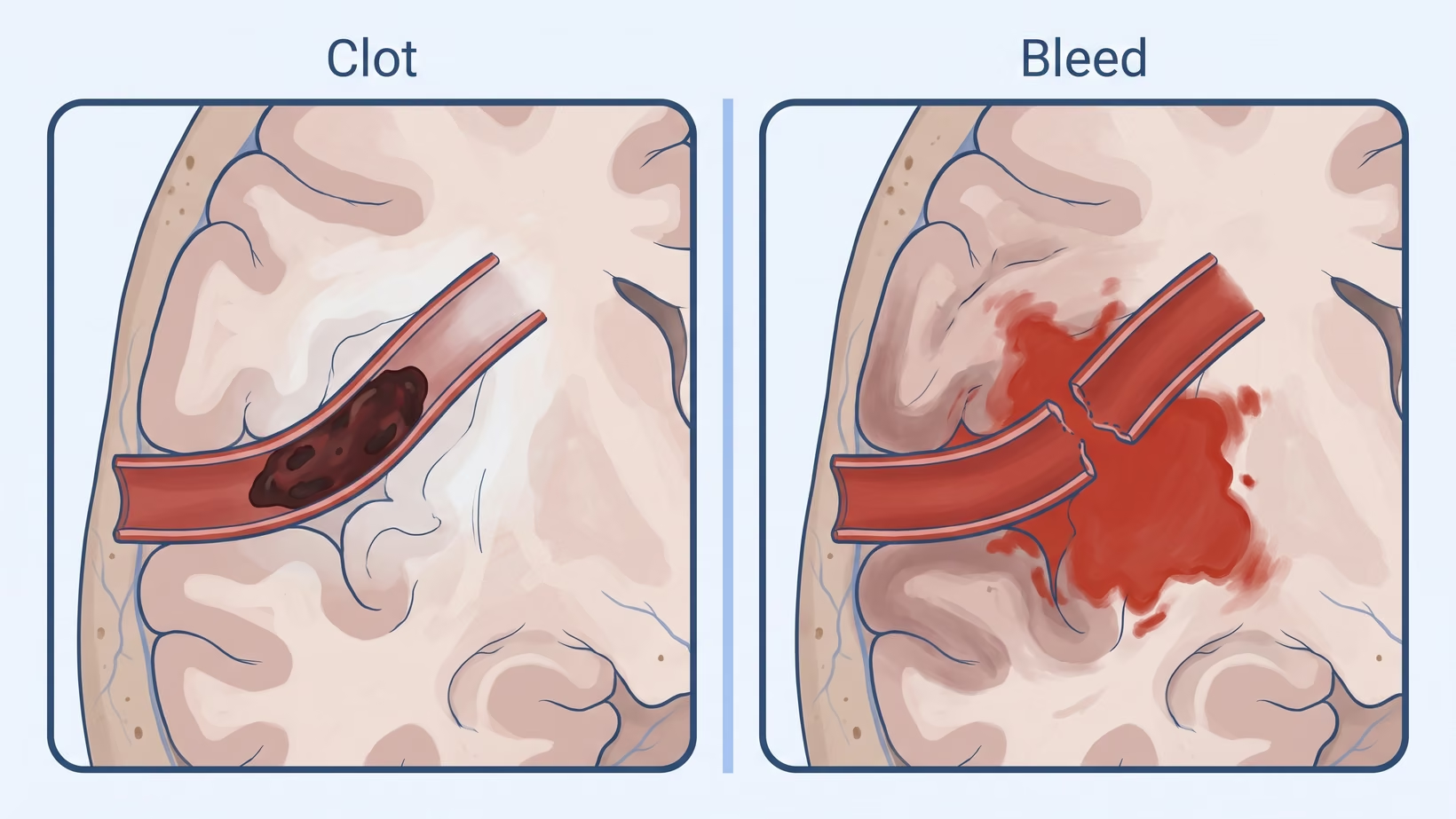
A hemorrhagic stroke is a stroke caused by bleeding. Blood escapes from a vessel into the brain, or into the space between the brain and its coverings. In the other kind of stroke, an ischemic stroke, a clot blocks the blood flow instead. Both kinds start suddenly, and both need the emergency room.
Every year, about 3.4 million people worldwide have a stroke from bleeding inside the brain that is not caused by an injury. Doctors call this a spontaneous bleed, meaning it happened on its own.
Bleeding after a head injury is a different problem, and it is also an emergency: go to the nearest emergency room now. The stroke guidelines on this page do not cover it.
The studies on this page did not report how common bleeding strokes are in the Philippines. The figures you will read here come from studies done worldwide or in other countries. They are not Philippine figures.
Seek care now
Warning signs: when is bleeding in the brain an emergency?
Any sudden sign of a stroke is an emergency, whether the cause is a bleed or a clot. Doctors outside hospital use FAST, the Face Arm Speech Test, which is a quick check of the face, arms and speech for a stroke. The test finds a possible stroke of either kind. Telling a bleed from a clot needs a brain scan.
Go to the nearest emergency room now if any of these start suddenly:
- One side of the face droops or looks uneven.
- One arm is weak or cannot be lifted.
- Speech is slurred, confused or lost.
- A severe headache starts suddenly.
- The person becomes drowsy or hard to wake.
Tell the emergency team at once if the person:
- Takes a blood thinner (anticoagulant), or is known to bleed easily.
- Is drowsy or hard to wake.
- Has signs that get worse, or come and go.
- Had a severe headache when the signs began.
- Has a stiff neck or a fever.
What should the family do right now?
Take the person to the nearest emergency room now. Do not delay leaving to gather things; do what you can on the way:
- Write down the time the signs began.
- If nobody saw them begin, write down the last time the person was seen well.
- Bring all the person’s medicines, or a photo of each label.
- Tell the emergency team if the person takes a blood thinner.
- Bring any old scans and reports.
Do not wait at home to see if the signs pass. Do not try hilot, massage or herbal remedies first. The emergency team will ask when the signs began, so that time helps them plan treatment.
What is an intracerebral hemorrhage?
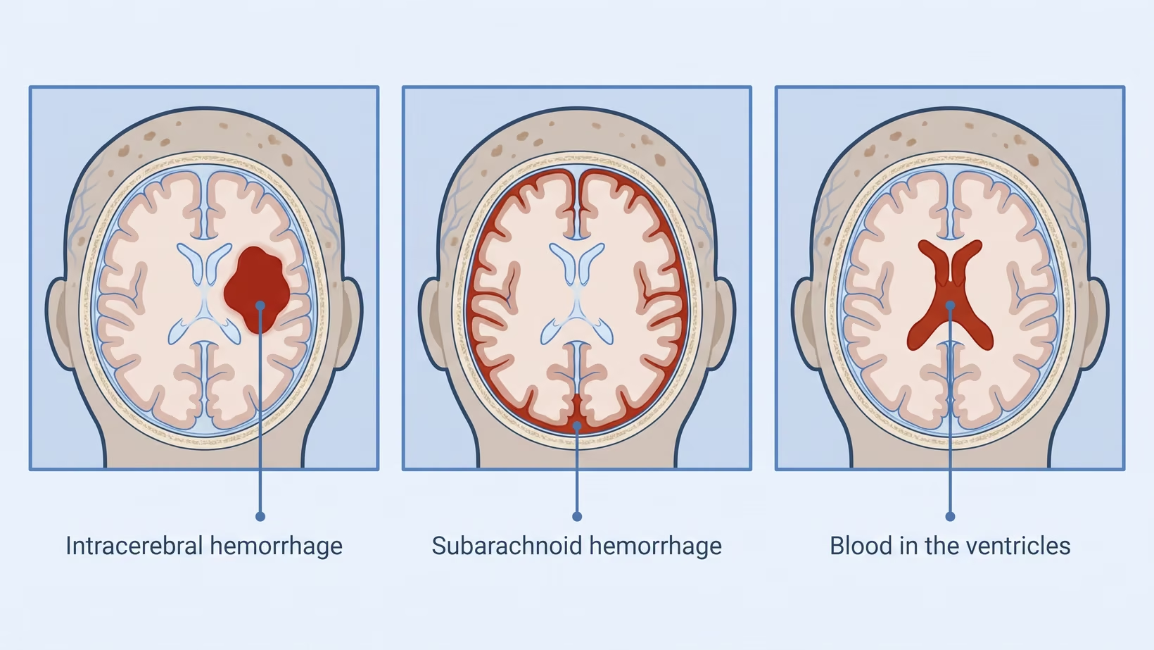
An intracerebral hemorrhage is bleeding inside the brain tissue itself. Intracerebral means inside the brain. The blood collects into a hematoma, meaning a pool of blood inside the brain. In 2019, about 28 in every 100 new strokes worldwide were this kind.
A bleed can grow after it starts. The European stroke guideline says most of this growth happens in the first 3 hours. Doctors try to limit the growth in the first hours, so the time the signs began matters.
Doctors measure the size of the hematoma on the scan, in milliliters (mL). Size is one of the things doctors weigh when they plan treatment. It is also linked to the outlook, which is explained further down this page.
What are the types of hemorrhagic stroke?
Doctors divide hemorrhagic strokes into two types by where the blood goes. An intracerebral hemorrhage is bleeding inside the brain. A subarachnoid hemorrhage is bleeding into the space between the brain and the thin membranes that cover it, like a lining around the brain.
A worldwide study counted every new stroke in 2019. Of every 100 new strokes:
- About 62 came from a clot (ischemic stroke).
- About 28 came from bleeding inside the brain (intracerebral hemorrhage).
- About 10 came from bleeding around the brain (subarachnoid hemorrhage).
These are worldwide estimates. The study did not report the split for the Philippines.
Blood from a bleed inside the brain can also spread into the ventricles. These are the fluid-filled spaces deep inside the brain. Sometimes fluid then builds up, a problem called hydrocephalus, and the person becomes less awake.
When that happens, the European guideline recommends a drain into those spaces, called an external ventricular drain, to reduce deaths. This recommendation comes from the agreement of the guideline’s experts, not from a trial.
What causes a hemorrhagic stroke?
The European guideline names getting older and high blood pressure among the main causes of a bleed inside the brain. It does not rank them or say how often each one is the cause.
A large study in 32 countries, called INTERSTROKE, compared people who had a stroke with people who had not. High blood pressure was more strongly linked with bleeding strokes than with clot strokes. Smoking, diabetes, heart problems and proteins that carry cholesterol were more strongly linked with clot strokes.
The study looked at risk factors that people may be able to change. Together they were linked with about 87 in 100 of the risk of bleeding strokes across the population. This is a share of risk for a whole population, not one person’s chance.
These are links found in a study. They do not prove that one factor caused one person’s bleed. A burst aneurysm, a weak spot in an artery that bulges like a small balloon, causes a different kind of bleed, around the brain.
Bleeding in the brain in babies, children and young adults
Bleeding in the brain also happens in babies, children and young adults. The guidelines on this page were written for adults, and they did not report causes or figures for children.
In younger adults, a bleed from a burst aneurysm is one cause to know. About half of the people with this kind of bleed are under 50.
If a baby or child suddenly becomes drowsy or hard to wake, or changes suddenly in any other way, go to the nearest emergency room now. Afterwards, ask the pediatrician or the neurosurgeon what may have caused the bleed. I treat children with bleeding in the brain at the Philippine Children’s Medical Center (PCMC) in Quezon City.
Where in the brain did it bleed, and why does that matter?
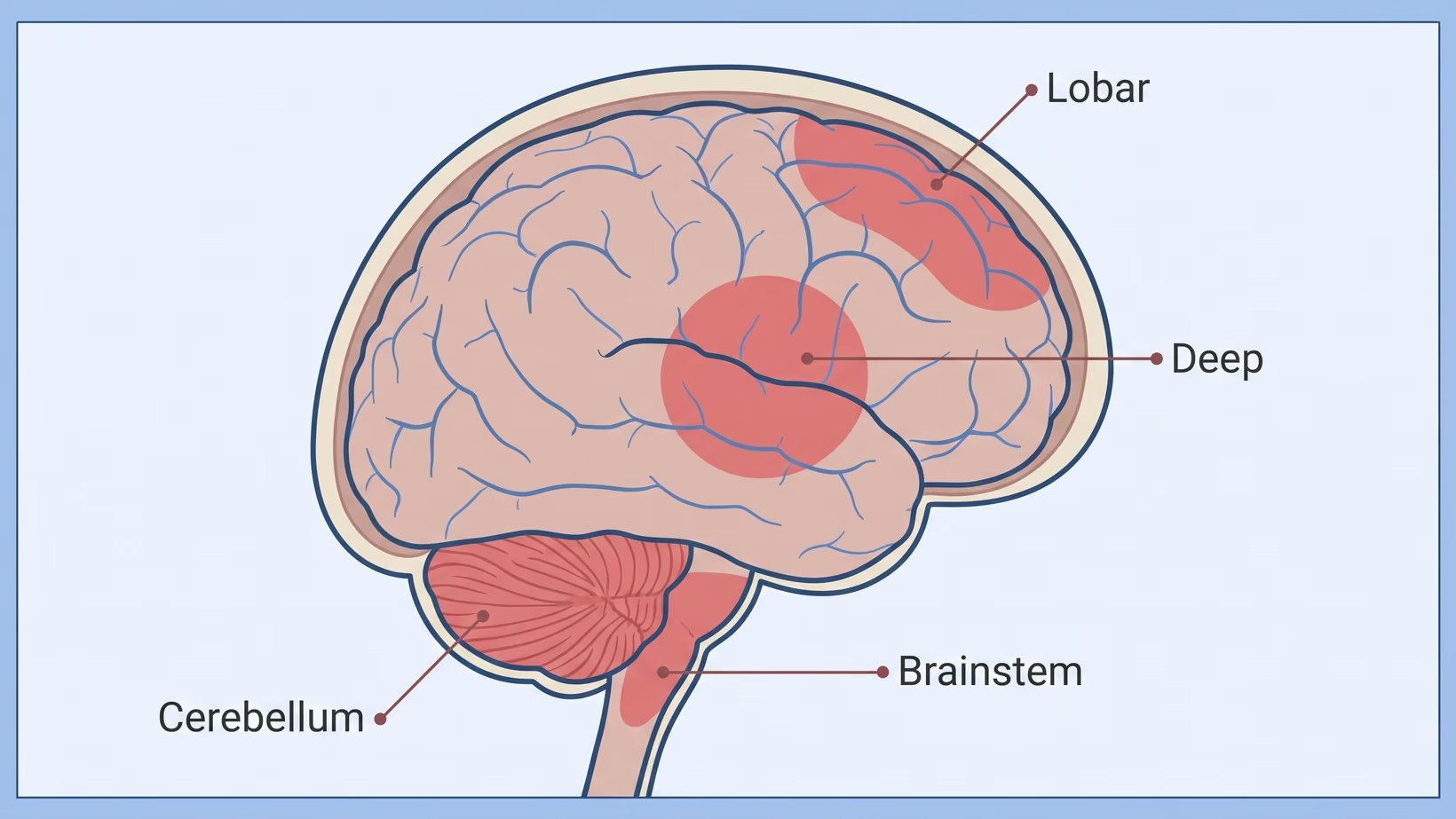
Where the bleed sits changes what doctors look for and whether surgery may help. Doctors describe the place of the bleed in these terms:
- Deep: in the basal ganglia or thalamus, groups of nerve cells deep in the middle of the brain.
- Lobar: in one of the lobes, the large outer parts of the brain.
- Cerebellum: at the back of the head, below the main part of the brain.
- Brainstem: the stalk that joins the brain to the spinal cord.
A bleed in the cerebellum sits in a small space. The European guideline says it is often linked with pressure on the brainstem, fluid build-up (hydrocephalus) and herniation, meaning brain tissue pushed out of its normal place.
For a cerebellar bleed above a certain size on the scan, the guideline suggests surgery to remove the blood, to improve survival. It is a weak suggestion. Recovery of function was similar with or without surgery.
For a lobar or cerebellar bleed in a younger person with no history of high blood pressure, doctors look further for a cause. They do this with scans of the blood vessels.
Why is a brain bleed treated differently from a clot?
A bleed and a clot are treated differently, so doctors find out which one it is before treatment starts. For a stroke caused by a clot blocking blood flow, doctors may give medicine that dissolves the clot. That medicine, and some other medicines for a clot stroke, are given only after a brain scan has shown there is no bleeding.
Blood pressure is another difference. The European guideline says to avoid lowering blood pressure before doctors know the type of stroke.
For a bleed, treatment aims to stop the bleed from growing and to protect the brain around it.
How do doctors tell a bleed from a clot?
Doctors tell a bleed from a clot with a brain scan. The UK stroke guideline says to do a plain CT scan, meaning one without dye, straight away in some patients. These include people on a blood thinner, people who are drowsy, and people who had a severe headache when the signs began.
Some patients then need a scan of the blood vessels. This can be a CT angiogram (a CT scan with dye), an MRI, or a catheter angiogram, where dye is passed into the arteries through a thin tube.
What happens in the first hours in hospital?
In the first hours, doctors try to stop the bleed from growing and watch for any worsening.
For a small or moderate bleed, the European guideline suggests lowering the top blood pressure number within 6 hours of the first signs. This aims to limit growth of the bleed. In the guideline’s review of trials, it had no clear effect on recovery or deaths at 3 months.
If the person takes warfarin, a blood thinner, the UK guideline says to bring clotting back to normal as soon as possible. Doctors do this with medicines that replace clotting factors, and vitamin K.
A patient who does not need intensive care should go to a stroke unit, a ward run by a stroke team. The European guideline recommends this to lower the risk of death or of needing others for daily care. Neurosurgery or stroke specialists watch for worsening and repeat the scan straight away when needed.
When does a brain bleed need surgery?
Whether a brain bleed needs surgery depends on where it is, its size and the person’s condition. For bleeds in the upper part of the brain, the European guideline also weighs timing, the method and the surgeon’s complication rate. This suggestion is weak and based on very low quality evidence.
In the STICH trial, early open surgery for upper brain bleeds gave a good result in about 26 in 100 patients. With medical treatment first, it was about 24 in 100. The trial found no overall benefit from early surgery.
In the ENRICH trial, early removal of the blood through a small opening gave better function at 6 months. By 30 days, about 9 in 100 had died in the surgery group and 18 in 100 in the other group. The benefit appeared to come from lobar bleeds.
What if the bleed came from an aneurysm?
When an aneurysm bursts, blood leaks into the space between the brain and its coverings. This is an aneurysmal subarachnoid hemorrhage, which needs care within the first 24 hours.
This type of stroke often happens at a relatively young age. About half of the people affected are under 50, and roughly two in three are women.
A burst aneurysm that is not treated bleeds again in about 40 in 100 patients. The European guideline says it should ideally be treated within 24 hours, when possible. A team with at least one neurosurgeon and one neurointerventionalist, a doctor who treats blood vessels through a thin tube in the arteries, decides together.
Other problems can follow. Reduced blood flow to the brain causes symptoms in about 20 in 100 patients. Fluid builds up in the brain in about 25 in 100.
Outcomes are often poor. About 40 in 100 patients die within 3 months. Roughly one in three of all patients regain independence in daily activities. These are European figures, not Philippine ones.
What is the outlook after bleeding in the brain?
The outlook after bleeding inside the brain is serious, and the figures differ widely between studies. In a review of population studies published from 1980 to 2008, a median of about 40 in 100 people died within one month. Across the studies this ranged from 13 to 61 in 100. In Japan it was lower, about 17 in 100.
Six of those studies measured how many people could live on their own afterwards. The figure was between 12 and 39 in 100.
The size of the bleed matters. The European guideline reports these deaths by size on the scan:
- A bleed of 60 mL (about four tablespoons) or more: over 90 in 100.
- A bleed of 30 mL (about two tablespoons) or more: about 30 to 55 in 100.
- A bleed under 30 mL: up to 20 in 100.
These are figures for groups. None of them tells you what will happen to one person. The guideline suggests that scores such as the ICH score should inform a person’s outlook, and not decide it alone.
The guideline also suggests that treatment should not be limited as a general policy in the first 24 hours. The studies above were mostly done outside the Philippines, and they did not report Philippine survival.
What to do now
When to refer a brain bleed: what the neurosurgeon needs
Refer early. The European guideline names patients such as these for an early neurosurgical consult: a large bleed, bleeding into the ventricles, or a bleed in the back of the brain that is taking up space.
The guideline gives no size cut-off for “large”. When to call the neurosurgeon about a brain bleed also depends on the points below.
- Cerebellar hematoma over 15 mL: the guideline suggests surgery to remove the blood, to improve survival. This is weak, and function was similar either way.
- Intraventricular extension with hydrocephalus lowering consciousness: an external ventricular drain is recommended to reduce deaths, by expert consensus.
- Younger patient, lobar or cerebellar bleed, no history of hypertension: CT angiography, MRI or MR angiography, or catheter angiography.
- On warfarin with a raised INR: NICE advises reversal with prothrombin complex concentrate and vitamin K, as soon as possible.
- Hematoma under 30 mL: the guideline suggests a top (systolic) blood pressure below 140 mmHg within 6 hours of onset, to limit expansion. It showed no clear effect on function or death at 3 months.
Send these with the referral:
- The CT images on a disc or an image link, and the report.
- The time last known well, and the time of the scan.
- The GCS score and pupils, and any change since arrival.
- The blood pressure readings and what was given.
- All medicines, with any blood thinner and the time of the last dose.
- The INR and platelet count.
If you are a family member and the person is still in hospital, ask the treating team about a neurosurgical consult. After discharge, book a consultation, or ask your doctor to refer you. If new weakness, a severe headache or drowsiness appears at home, go to the nearest emergency room now; do not wait for the consultation. Bring the scans on a disc, the reports and a list of all medicines.
You may also see me, or your doctor may refer the patient to me:
- Adults: Lung Center of the Philippines, Room 1101, Quezon Avenue, Quezon City, Tuesday and Thursday from 2 PM.
- Children: Philippine Children’s Medical Center, Room 22, Quezon Avenue corner Agham Road, Diliman, Quezon City, Wednesday from 2 PM.
- If the trip to Quezon City is not practical, I also see patients online. Book it on the consultation page.
Frequently asked questions
Is a brain bleed the same as a stroke?
A brain bleed that happens on its own, not from an injury, is a type of stroke called a hemorrhagic stroke. About 3.4 million people worldwide have this kind of bleed inside the brain each year. Bleeding after a head injury is a different problem, but it is also an emergency that needs the nearest emergency room now. The stroke guidelines on this page do not cover it.
Can bleeding in the brain happen again?
Another stroke can happen after bleeding in the brain. The European guideline strongly recommends blood pressure control after a brain bleed, to lower the risk of a later stroke. It does not give the chance of a second bleed. A burst aneurysm that is not treated bleeds again in about 40 in 100 patients.
Why does a blood thinner matter in a brain bleed?
A blood thinner matters because doctors scan straight away when a person on one has signs of a stroke. For someone on warfarin, the UK guideline says to bring clotting back to normal as soon as possible. Do not stop a blood thinner on your own. Ask the doctor who prescribed it.
How much does treatment cost, and does PhilHealth help?
The cost of treating bleeding in the brain depends on the hospital, the length of stay and whether surgery is needed. The sources on this page give no Philippine cost or PhilHealth figure. Ask the hospital billing office for an estimate, and ask the PhilHealth desk what your membership covers.
See sources
- Steiner T, Purrucker JC, Aguiar de Sousa D, et al; ESO and EANS. European Stroke Organisation (ESO) and European Association of Neurosurgical Societies (EANS) guideline on stroke due to spontaneous intracerebral haemorrhage. European Stroke Journal. 2025. PMID 40401775; doi:10.1177/23969873251340815.
- GBD 2019 Stroke Collaborators. Global, regional, and national burden of stroke and its risk factors, 1990-2019: a systematic analysis for the Global Burden of Disease Study 2019. Lancet Neurology. 2021. PMID 34487721; doi:10.1016/S1474-4422(21)00252-0.
- O’Donnell MJ, et al; INTERSTROKE investigators. Global and regional effects of potentially modifiable risk factors associated with acute stroke in 32 countries (INTERSTROKE): a case-control study. Lancet. 2016. PMID 27431356; doi:10.1016/S0140-6736(16)30506-2.
- National Institute for Health and Care Excellence. Stroke and transient ischaemic attack in over 16s: diagnosis and initial management (NICE guideline NG128). 2019, updated 2022. https://www.nice.org.uk/guidance/ng128
- Mendelow AD, Gregson BA, et al; STICH investigators. Early surgery versus initial conservative treatment in patients with spontaneous supratentorial intracerebral haematomas in the International Surgical Trial in Intracerebral Haemorrhage (STICH): a randomised trial. Lancet. 2005. PMID 15680453; doi:10.1016/S0140-6736(05)17826-X.
- Pradilla G, et al; ENRICH investigators. Trial of Early Minimally Invasive Removal of Intracerebral Hemorrhage. New England Journal of Medicine. 2024. PMID 38598795; doi:10.1056/NEJMoa2308440.
- Vergouwen MDI, Ramos-Pachon A, Terecoasa EO, et al; ESO, EANS and ESMINT. European Stroke Organisation (ESO), European Association of Neurosurgical Societies (EANS) and European Society for Minimally Invasive Neurological Therapy (ESMINT) guideline on aneurysmal subarachnoid haemorrhage. European Stroke Journal. 2026. PMID 42095754; doi:10.1093/esj/aakag043.
- van Asch CJ, et al. Incidence, case fatality, and functional outcome of intracerebral haemorrhage over time, according to age, sex, and ethnic origin: a systematic review and meta-analysis. Lancet Neurology. 2010. PMID 20056489; doi:10.1016/S1474-4422(09)70340-0.