

Midline shift means the brain has been pushed across the middle of the skull by something taking up space on one side: blood, swelling or a tumor. A radiologist measures it in millimeters. After an injury, or if the person is getting worse, go to the emergency room now; otherwise a neurosurgeon should see the scan the same day.
What is midline shift on a CT scan?
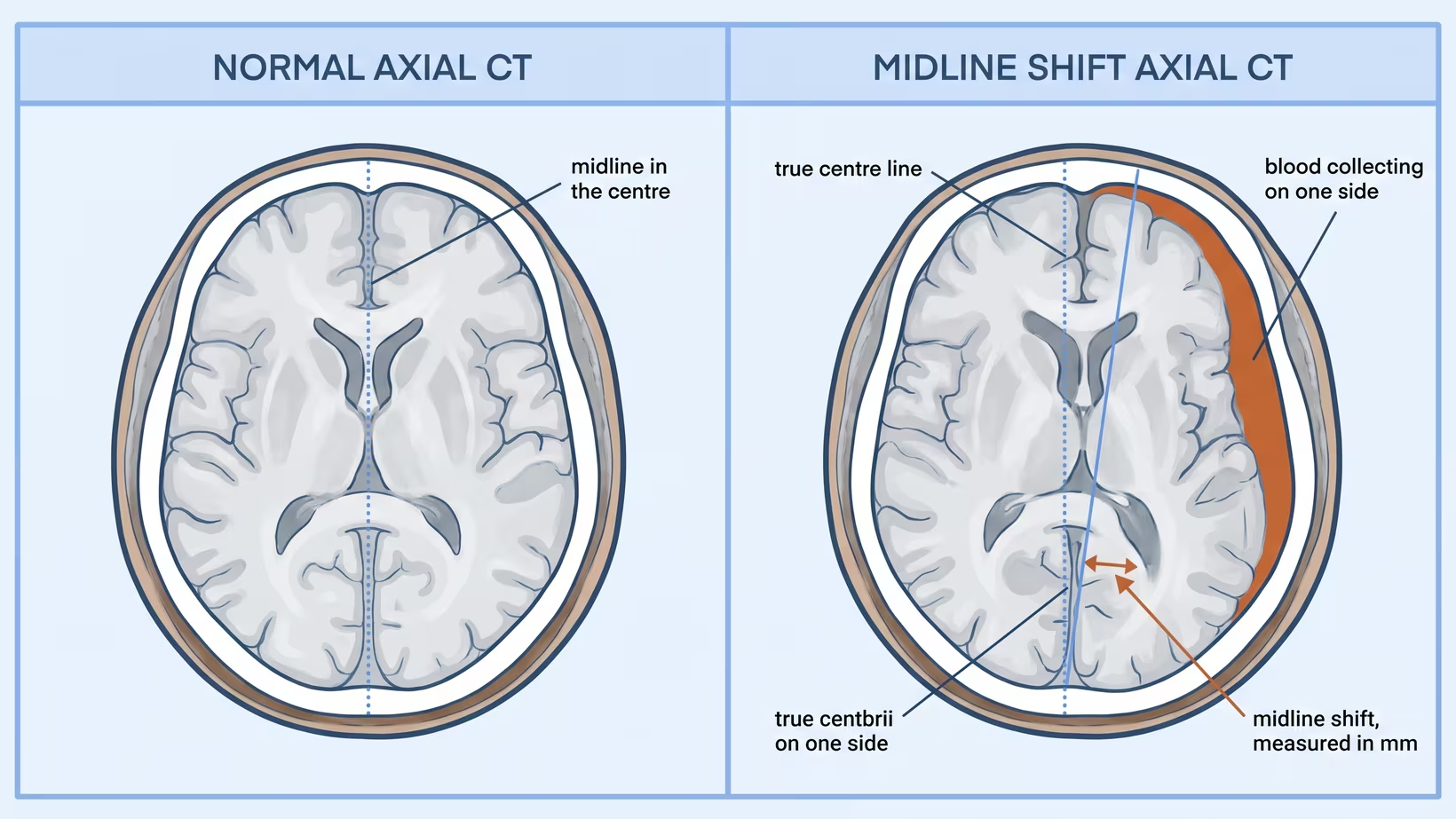
Midline shift is the displacement of the brain’s midline structures across the centre of the skull. It is reported in millimetres, and that figure comes from the radiologist’s measurement on the axial CT images.
On a scan with nothing pushing on it, the structures that sit between the two halves of the brain line up with the middle of the skull. When something on one side takes up room, those structures are pushed across. The distance they have moved is the shift.
The word shift describes a position, not a diagnosis. It tells you that something inside the skull is occupying space that the brain was using. It does not tell you what that something is. A report may read “mass effect with 8 mm of midline shift” or “no midline shift”; both phrases are describing the same measurement.
What does it mean when the brain is pushed to one side?
When the brain is pushed to one side, there is no longer room inside the skull for everything in it, and the brain has moved because it had nowhere else to go. The skull does not expand in an adult, so extra volume on one side is paid for by the brain on the other side.
For a family, this is the sentence that matters: the shift shows how far the brain has been pushed, not how much blood there is.
After a head injury, midline shift is one of the CT findings that carries information about how a patient is likely to do. In models for predicting outcome, built from 10,008 patients in the MRC CRASH trial and tested in a further 8,509, midline shift sat alongside age, the Glasgow Coma Scale (a score of how awake the patient is), pupil reactivity and other CT features.
It is read together with the patient’s condition and never as a verdict on its own. No millimetre figure predicts what will happen to one individual, and those models performed less well when tested outside the populations that built them.
How does a subdural hematoma cause midline shift?
A subdural hematoma causes midline shift by taking up space on one side of the skull, so the brain beneath it is pushed across the middle. A subdural hematoma is blood collecting between the surface of the brain and the tough membrane that lines the skull. It spreads over one hemisphere (one half of the brain), and every millilitre of it occupies space the brain was already using.
Because the collection is on one side, the pressure it creates is one-sided too. The brain under it is pushed away from the skull and across the middle. A thin layer of blood can sit alongside a large shift. When the shift is much bigger than the clot is thick, the brain underneath is usually swollen as well, and surgeons treat that as a more serious picture, not a milder one.
A subdural hematoma usually reaches the scanner by one of two routes. One is a sudden bleed after a hard impact, which is an acute subdural hematoma: a fall down the stairs, a motorcycle crash, a blow to the head. The other is a slow collection in an older adult, often weeks after a bump that seemed minor at the time.
What is mass effect, and why does it matter?
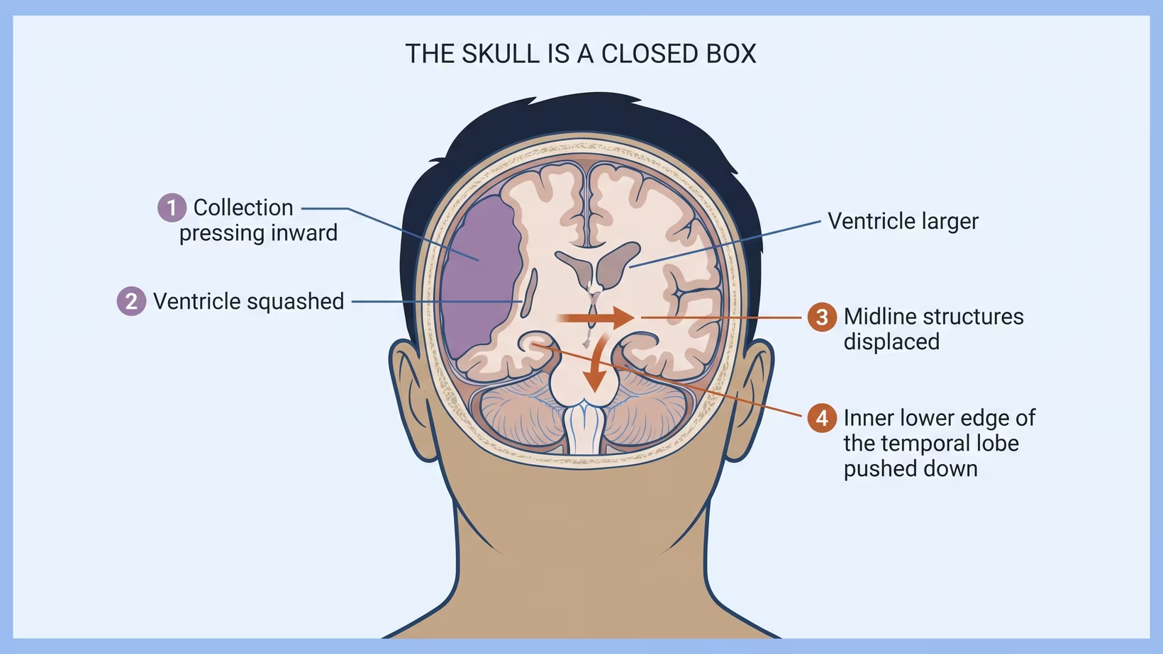
Mass effect is the name for what the extra volume does to everything around it. The clot is the cause; mass effect is the damage, and it is what the surgeon is actually treating.
Follow it in order. The collection sits over one hemisphere. The ventricle on that side, one of the fluid spaces inside the brain, is squashed flat, while the one on the other side may enlarge. The midline structures are pushed across. The inner edge of the temporal lobe, low down on the affected side, is pressed towards the narrow opening beside the brainstem.
Pressure inside the skull is treated as a problem in its own right. The Brain Trauma Foundation recommends treating intracranial pressure, meaning the pressure inside the skull, above 22 mm Hg, because values above that level go with more deaths, and it states that the pressure reading is used together with the clinical picture and the CT findings rather than on its own. That is the reasoning behind a phrase families often hear in the unit: we are treating the pressure and the displacement, not the blood.
The older guideline for operating on an acute subdural hematoma uses 20 mm Hg for a comatose patient with a small clot, which is the figure in the referral list below.
What do the pupils tell you about the shift?
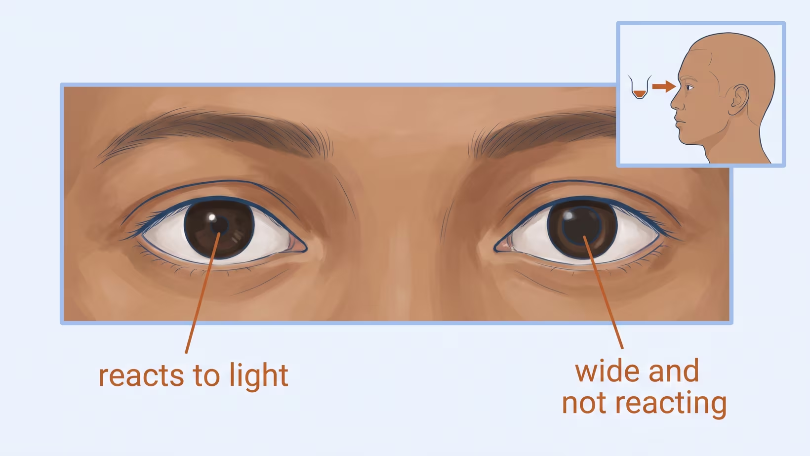
A pupil that becomes wider than the other, or stops reacting to light, can mean the shifting brain is pressing on the nerve that controls it, and it makes the situation more urgent. Pupils can be looked at without a scanner, which is why they are checked again and again.
In the guideline written for a comatose patient with a traumatic acute subdural hematoma, pupils that are unequal, or fixed and dilated, are one of the findings that tips the decision towards surgery even when the clot is thin and the shift is under 5 mm.
The anatomy is the reason. The nerve that makes a pupil constrict runs close to that inner edge of the temporal lobe, the part pressed towards the brainstem as the midline moves.
A pupil is not a substitute for the scan, and an unchanged pupil is not reassurance on its own. It is a finding that changes the urgency when it appears.
For families: you do not need to examine anyone’s pupils yourself. If you notice that one of your relative’s pupils looks wider than the other, or that they have become harder to wake, tell the nurse straight away rather than waiting for the next round.
If the person is at home and becomes harder to wake, is getting worse in any other way, or has one pupil wider than the other, even after a minor head injury weeks ago, take them to the nearest emergency room now. Do not wait for a clinic appointment.
Seek care now
When to refer, and when it cannot wait
For the doctor who has the scan:
New midline shift after a head injury, or with a patient who is getting worse, is a phone call to a neurosurgeon on the same shift it is seen, not a referral letter. Once an acute subdural hematoma meets the criteria for surgery, the guideline says the evacuation should be performed as soon as possible.
Call now, before the patient is fully worked up, if any of these is present:
- A traumatic acute subdural hematoma with midline shift greater than 5 mm, or a clot thicker than 10 mm, at any level of consciousness
- A drop of two or more points on the Glasgow Coma Scale between the injury and now
- Pupils that are unequal, or fixed and dilated
- Intracranial pressure above 20 mm Hg, where it is being monitored
Have this ready when you call: the CT images and the written report, the time and mechanism of injury, the Glasgow Coma Scale then and now, the pupil findings, and the medicine list, with any blood thinners named: anticoagulants (which stop clotting factors from working) and antiplatelets (which stop platelets from sticking together). Say plainly whether the patient is deteriorating.
Distance is part of the decision here. A 2023 survey counted 174 neurosurgeons in the Philippines, roughly one for every 600,000 people, with 35 provinces having none at all, and burr holes (small holes drilled through the skull) or craniotomy (opening a section of the skull) for head injury among the most common operations they perform.
If the nearest service is a transfer away, the call is made while the patient is still being stabilised, not after. Expect the neurosurgeon to want the images themselves and to advise on what is actually done about the clot before the patient moves.
How many millimetres of midline shift matter?
For a traumatic acute subdural hematoma, the figure in the guideline is 5 mm. The Brain Trauma Foundation and Congress of Neurological Surgeons recommend that a clot thicker than 10 mm, or a midline shift greater than 5 mm on CT, be taken out by surgery whatever the Glasgow Coma Scale score.
Two things about that sentence are easy to misread. First, it is a recommendation to operate, not a measurement of danger: 4 mm is not a safe number and 6 mm is very bad. Second, it was written for an acute traumatic bleed.
It does not automatically apply to a chronic collection, to swelling after a stroke, or to a tumour. Those displace the midline for different reasons and over different timescales, and they are assessed on their own terms.
How the shift is actually measured on the scan
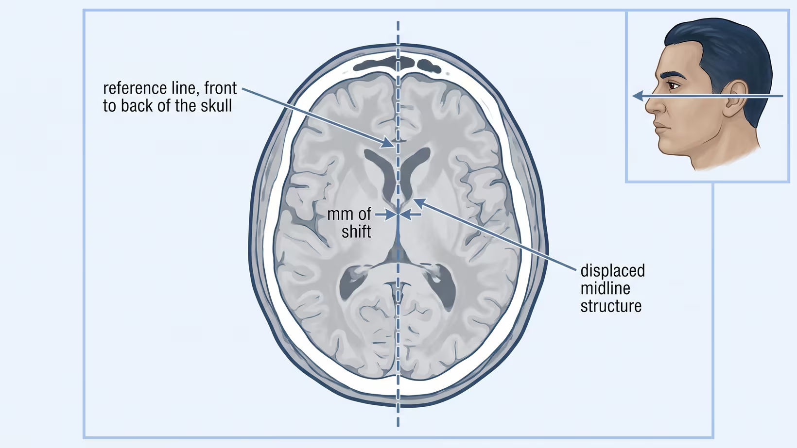
The measurement is a distance taken on one axial slice, at the level of the foramen of Monro, the small channel that links the fluid spaces on each side to the centre of the brain. The radiologist draws a reference line down the true midline of the skull, from front to back, finds the structure that should sit on that line between the two ventricles, and measures the perpendicular distance from the line to where the structure actually lies.
That distance, in millimetres, is the shift. Because it is a single measurement on a single slice, it depends on the level chosen and on who takes it. What the team follows is the direction of travel between scans, not one figure in isolation.
Why operate for the shift, not the size of the clot?
Surgeons operate for the shift because it shows what the clot is doing to the brain. In the guideline for a traumatic acute subdural hematoma, either a clot thicker than 10 mm or a shift of more than 5 mm meets the recommendation for surgery, whatever the patient’s Glasgow Coma Scale score, and the neurosurgeon still weighs it against the patient in front of them.
The reverse also holds. In a comatose patient whose clot is under 10 mm thick and whose shift is under 5 mm, the same guideline recommends surgery if the Glasgow Coma Scale dropped by two or more points between the injury and arrival at hospital, or the pupils are unequal or fixed and dilated, or the intracranial pressure is above 20 mm Hg. The scan is never read alone.
Making room is not free. When pressure will not come down with medical treatment, a different operation, removing part of the skull, was tested against continued medical care in a randomised trial of 408 patients: it lowered the death rate at six months, but left more survivors in a vegetative state or severely disabled. The operation has real costs as well as benefits, and that is why no millimetre figure makes the decision automatic.
Does the shift come back to the middle after the clot is removed?
Removing what is taking up the space is what allows the midline to move back towards the centre. How much of that happens, and how quickly, depends on what was there and how long it had been there.
A follow-up scan checks the response to treatment, and how far the midline returns is not something anyone can promise in advance. Whether the person recovers is a separate question from whether the shift is restored, and what the outlook depends on is more than the millimetres shifted.
Does it matter how fast the shift developed?
How fast midline shift developed matters a great deal, and it is usually the first thing the surgeon asks about. The same number of millimetres does not carry the same meaning in a bleed that formed in an hour and in a collection that built over weeks.
The 5 mm figure belongs to the fast bleed. It was written for the traumatic acute subdural hematoma, not for slow collections, strokes or tumors, so a slowly grown collection with a similar shift is not read against it. That does not make a slow collection with shift safe to leave: it still needs a neurosurgeon’s review the same day, and sooner if the person is getting worse.
At the other end of the spectrum is a chronic subdural hematoma in an older adult, which the authors of a large trial describe as a common neurological disorder, especially common among older people. In that multicenter trial of 748 adults, the mean age was 74, and about 94 in 100 (94 percent) had the collection surgically drained during the same admission. Those were patients who already had symptoms, so it is not a rule that every chronic collection is operated on.
What else pushes the midline across besides a subdural hematoma?
Anything that takes up room on one side of the skull can do it: blood inside the brain as well as outside it, a large stroke that swells, a tumor, an abscess, or a collection of fluid rather than blood. The CT report describes the displacement; the rest of the scan says what caused it.
A stroke is worth spelling out, because swelling is treated on the same logic as a clot. In a pooled analysis of three randomised trials, surgery within 48 hours of a large stroke reduced the number of deaths.
This stroke is an infarction, meaning brain tissue that has died after losing its blood supply, and it takes up space in the skull. It was in the middle cerebral artery, a main artery supplying one side of the brain. The authors state that the decision has to be made for each patient individually.
Read that carefully before carrying it anywhere else. Those trials enrolled 93 patients aged 18 to 60 with one specific kind of stroke, and many of those who survived lived with moderately severe disability rather than returning to normal.
A slow-growing tumor such as a meningioma is the opposite situation: displacement that developed over months rather than hours, often found on a scan ordered for headaches, seizures or a slow change in the person, rather than after an injury.
Frequently asked questions
Is midline shift always an emergency?
Midline shift should be shown to a neurosurgeon the same day it is found. If it follows an injury or the person is getting worse, go to the nearest emergency room now. Whether it means an operation tonight depends on the cause, on how fast it developed, and on how the patient is: for a traumatic acute subdural hematoma, a shift of more than 5 mm meets the guideline criterion for evacuating the clot.
Can a person be awake and talking with midline shift?
Yes. For a traumatic acute subdural hematoma with more than 5 mm of shift, the guideline recommends surgery whatever the Glasgow Coma Scale score, so being awake does not mean the team will wait. Being awake is not evidence that the shift is small.
Can midline shift be seen on an X-ray or an ultrasound?
Not on a skull X-ray, which shows bone rather than the position of the brain. Ultrasound through the skull can estimate it in some intensive care units, and MRI shows it, but a CT scan of the head is the usual test. If the question has been raised and no CT has been done, the CT is the test that answers it.
Will the CT scan need to be repeated?
Often, yes, because a repeat scan is how the team sees whether the shift is increasing or settling. There is no fixed interval to expect: it is ordered on the basis of how the patient is doing, and a change in alertness is a reason to bring it forward.
Should we ask for a copy of the scan and the report?
Yes. Ask for the images on a disc or a drive and for the written report, because the next doctor will want to look at the scan itself rather than a description of it. Bring the medicine list, blood thinners especially, and be ready to say when the injury happened and how the person has changed since.
Does midline shift mean permanent brain damage?
Not by itself. Midline shift is one of several findings that carry information about outcome after a head injury, alongside age, the level of consciousness and the pupil response, and no millimetre figure predicts what happens to one person. Ask the team treating the patient what this particular scan means in this particular case.
See sources
- Bullock MR et al, for the Surgical Management of Traumatic Brain Injury Author Group. Surgical management of acute subdural hematomas. Neurosurgery (Guidelines for the Surgical Management of Traumatic Brain Injury, Brain Trauma Foundation and Congress of Neurological Surgeons). 2006. PMID 16710968.
- Carney N, Totten AM et al, Brain Trauma Foundation. Guidelines for the Management of Severe Traumatic Brain Injury, Fourth Edition. Neurosurgery. 2017. PMID 27654000. doi:10.1227/NEU.0000000000001432
- MRC CRASH Trial Collaborators (Perel P, Arango M et al). Predicting outcome after traumatic brain injury: practical prognostic models based on large cohort of international patients. BMJ. 2008. PMID 18270239. doi:10.1136/bmj.39461.643438.25
- Hutchinson PJ et al, RESCUEicp Trial Collaborators. Trial of Decompressive Craniectomy for Traumatic Intracranial Hypertension. New England Journal of Medicine. 2016. PMID 27602507. doi:10.1056/NEJMoa1605215
- Vahedi K et al. Early decompressive surgery in malignant infarction of the middle cerebral artery: a pooled analysis of three randomised controlled trials. The Lancet Neurology. 2007. PMID 17303527. doi:10.1016/S1474-4422(07)70036-4
- Hutchinson PJ, Edlmann E, Bulters D et al, British Neurosurgical Trainee Research Collaborative. Trial of Dexamethasone for Chronic Subdural Hematoma. New England Journal of Medicine. 2020. PMID 33326713. doi:10.1056/NEJMoa2020473
- Baticulon RE, Lucena LLN, Gimenez MLA et al. The Neurosurgical Workforce of the Philippines. Neurosurgery. 2023. PMID 37931081. doi:10.1227/neu.0000000000002630