
A chronic subdural hematoma is a slow collection of blood and fluid on the surface of the brain. It happens mostly in older people, because the brain shrinks with age and stretches the veins under its outer covering. A small bump can tear one. Go to an emergency room now for drowsiness, weakness on one side or repeated vomiting.
What Does a Slow Bleed on the Brain Mean for an Elderly Patient?
It means blood has collected in a thin layer over the surface of the brain and has been slowly taking up space there for weeks. There is no spare room inside the skull, so even a small collection eventually presses on the brain.
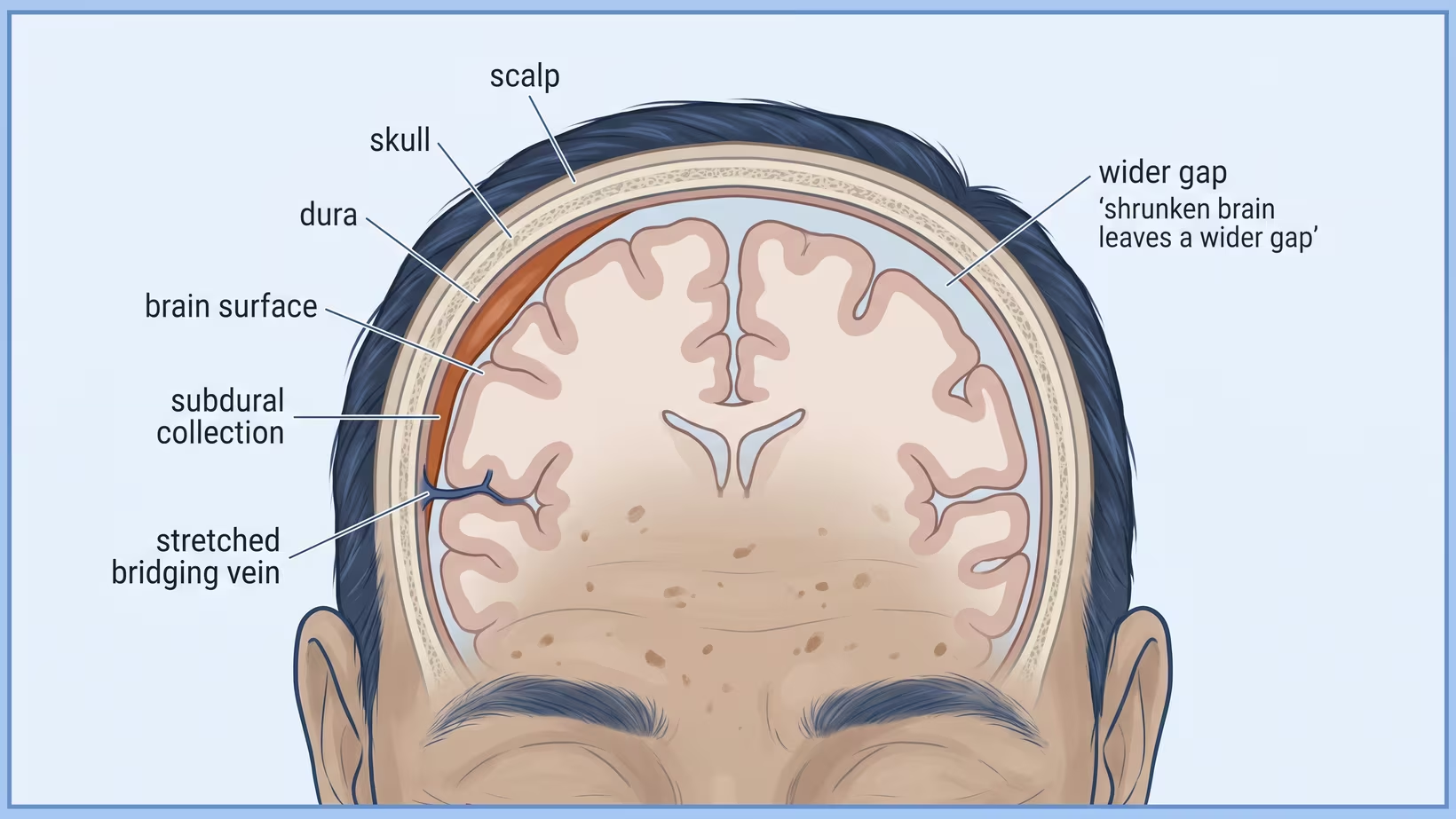
How serious it is depends on how much space the collection is taking up and on how your father is doing, not on the word hematoma. The collection lies between the dura, the tough outer layer that covers the brain, and the thin arachnoid layer under it. It is enclosed in its own thin lining, like a pouch. The lining can bleed and leak, and this keeps the collection growing.
Slow Is Not the Same as Harmless
Chronic refers to time, not to severity. A collection is usually called chronic after three weeks. A chronic collection is not a milder form of the emergency. It is blood collecting in the same space, only much more slowly. Because it grows slowly, a patient can have it for a month while the family thinks something else is wrong. The acute type, the fast bleed, which behaves completely differently, causes symptoms within hours.
Why Does an Old Brain Bleed After Such a Small Bump?
Because the brain shrinks with age. This leaves a wider gap between the brain and the skull. The bridging veins, which run across that gap from the brain to the dura, are pulled tight. A stretched vein tears more easily than a loose one, especially when the head moves suddenly and the brain shifts inside the skull.
The known risk factors are older age, being male, and taking antiplatelet or anticoagulant drugs, which are blood thinners. How often it happens differs widely between populations, from 1.72 to 20.6 cases per 100,000 people each year.
What If Nobody Remembers a Fall at All?
That is common, and it does not make the diagnosis less likely. The medical literature is explicit that a chronic subdural hematoma usually follows minor trauma but that the absence of remembered trauma is common and should not lower diagnostic suspicion.
Do not blame yourself if you cannot remember or suspect a patient of hiding the trauma. The patient may genuinely have not noticed. A head that taps a doorframe or a jeepney roof, a stumble in the bathroom that ended with a hand on the wall, a grandchild’s elbow: none of these gets remembered a month later, and none of them needs to be.
Why the Injury Is Often Missed
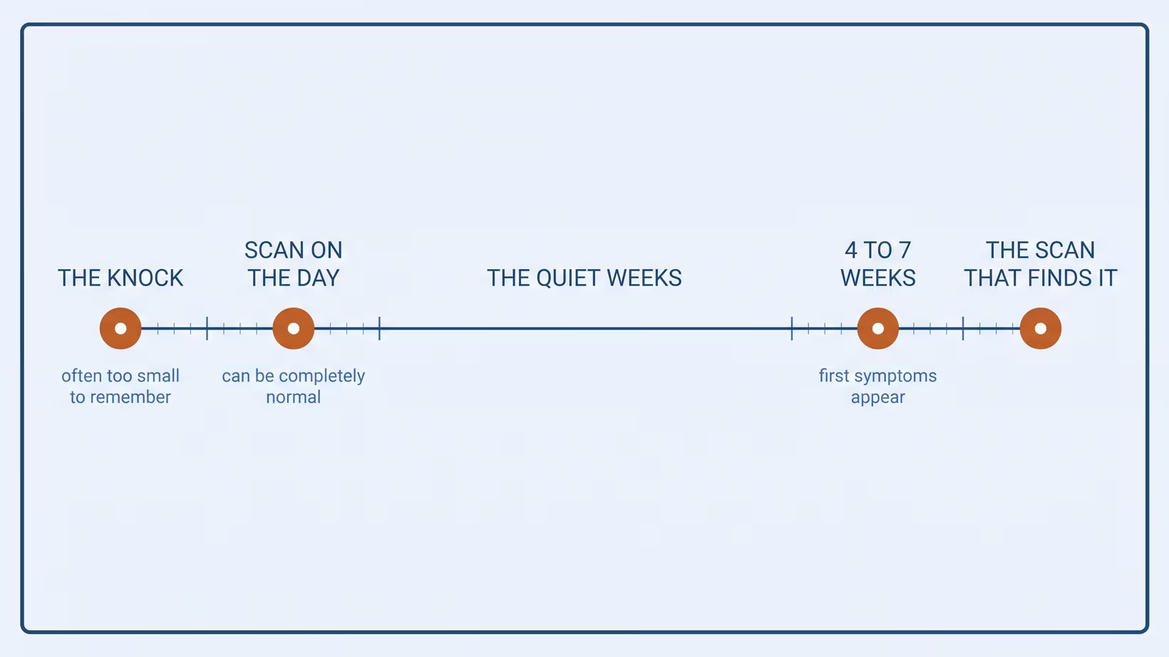
It is missed for two reasons. First, the original bump caused no symptoms, so nobody wrote it down. Second, a scan taken at the time of the injury can be completely normal, with no sign of hemorrhage (bleeding). The patient can still develop a collection weeks to months later.
Seek care now
Which Signs Mean the Emergency Room Now?
A chronic collection usually present itself slowly, so the signs that matter are the ones that break that pattern. Take your parent to an emergency department now, not in the morning, for any of these.
- A sudden, severe headache, or a headache clearly worse than yesterday’s
- A drop in how awake he is: hard to wake, drowsy between sentences, or sleeping through the day
- New weakness or numbness on one side of the body, or a face that has begun to droop
- Speech that has become slurred, or trouble finding words
- Repeated vomiting, especially together with drowsiness
- A first seizure, or any jerking of the arms, legs or body
- Confusion that has come on over hours rather than weeks
- Any new head injury in someone taking a blood thinner, even if he seems fine
Antiplatelet and anticoagulant drugs are a known risk factor for this condition. A patient on a blood thinner who looks well on the evening of a fall can still develop a collection in the weeks after.
How Did the Doctor Find It?
Almost always, a plain CT scan of the head is diagnostic, it ordered because something about the patient is no longer normal. It is not hard to diagnose, once a scan is done, the difficulty is in deciding to scan at all.
The symptoms that lead to the scan are often milder than families expect. In older adults, walking often gets worse, and this is often a better clue than headache. Patients may also have repeated falls, slow thinking, loss of interest in things (apathy), or confusion that comes and goes. Others have a new, urgent need to urinate, mild trouble finding words, a clumsy hand, weakness on one side, or a drooping face.
Why a Plain CT Is Usually Enough
This kind of collection shows clearly on an ordinary head CT, which is fast and available in most Philippine hospitals with an emergency department. An MRI shows finer detail, but it is usually not the first test for this.
What the Words on the Report Mean
A report on this usually names three things: which side the collection is on, how thick it is in millimeters, and whether the brain has been pushed across the midline. The surgeon reads the third one first.
Ask for a copy, or photograph the report with your phone before you leave the ER. If your father is later seen by a second doctor, that piece of paper saves a repeat scan. In these cases, symptoms show up weeks after a bump on the head, and should prompt a repeat scan.
The Scan Says Brain Shrinkage. Is That the Same Problem?
No. Brain shrinkage, written on the report as cerebral atrophy or generalized volume loss, is the background condition that made the bleed possible. It is not the bleed, and it is not the reason your father is unwell today. The two findings often appear in the same paragraph of the report, so families often read them as one problem. The collection can be drained. The volume loss cannot, and does not need to be.
Atrophy Is the Setting, Not the Diagnosis
Some volume loss is expected in an ageing brain and is the very thing that widens the space and stretches the bridging veins, so this the part of the report describing the mechanism of the disease rather than a second disease. What it does not tell you is whether your father has dementia. Those are different questions answered by different assessments, and the confusion between them is common enough that we need to know how to differentiate it from dementia.
Why Does It Keep Growing for Weeks?
Because the body does not simply absorb the collection. A lining forms around it, and that lining bleeds and leaks. So the space keeps filling long after the torn vein has stopped bleeding. On average, it takes four to seven weeks after the injury before the collection causes symptoms.
What Are the Membranes, and Why Do They Bleed Again?
The membranes are the new linings the body grows around the collection, as part of the inflammation after the original injury. The outer one is full of fragile new blood vessels. These vessels leak tiny bleeds and fluid into the space inside the membranes. They are what keeps the collection growing.
The Cycle That Keeps It Going
This is a repeating cycle rather than a single bleed. Inflammation builds membranes. The membranes grow fragile vessels. The vessels bleed and leak, and that bleeding causes more inflammation.
Why This Is Also the Reason It Can Come Back
An operation empties the space. It does not remove the membranes, and the membranes are what produce the fluid. About 11 in 100 patients treated with surgery (11%) have a recurrence, meaning the collection comes back and needs another operation. This is why the follow-up scan must be done.
Who Should Be Referred, and How Fast?
In my practice, any patient with a symptomatic subdural collection on CT is discussed with a neurosurgeon on the day of the scan. How urgent it is depends on the neurological signs and how awake the patient is, not on the millimeters.
The common European rule for operating is a hematoma thicker than the skull bone. That is a threshold for operating, not a threshold for referring.
The Referral That Cannot Wait
Refer these patients now, by phone as well as by letter: a patient who is less awake, or whose alertness comes and goes; a new or worsening focal deficit, such as weakness of one side; a seizure; or a collection with significant midline shift. I treat these patients as emergencies from the moment they arrive, however old the blood looks on the scan.
The Referral That Can Be Booked
A patient who is stable, with mild symptoms and a modest collection, can be given a scheduled appointment instead of being transferred at midnight. In a group of 444 patients with mild to moderate symptoms, 114 were first managed without surgery. Of those 114, 49, about 43 in 100, later needed surgery.
Watching and waiting is a valid choice in selected patients, but the follow-up has to be scheduled and kept.
I see these booked referrals at my clinic at the Lung Center of the Philippines. A patient who lives far from Quezon City can be seen in an online consult instead.
What the Referring Doctor Should Send
Send the CT images, not only the report, with the items in the checklist below.A German group of 395 surgical patients found that stopping blood thinners was linked to complications from blood clots in about 9 in 100 of them (9.1%). The authors concluded that the decision has to be made carefully for each patient.
What Do the Studies Show About Outcomes?
Clinical trials in the last 20 years have improved the operation. But the surgical figures alone do not describe the year after the operation. After the first month, most deaths come from other illnesses, such as dementia and heart disease, not from the collection.
Surgery, and the Drain That Changed the Numbers
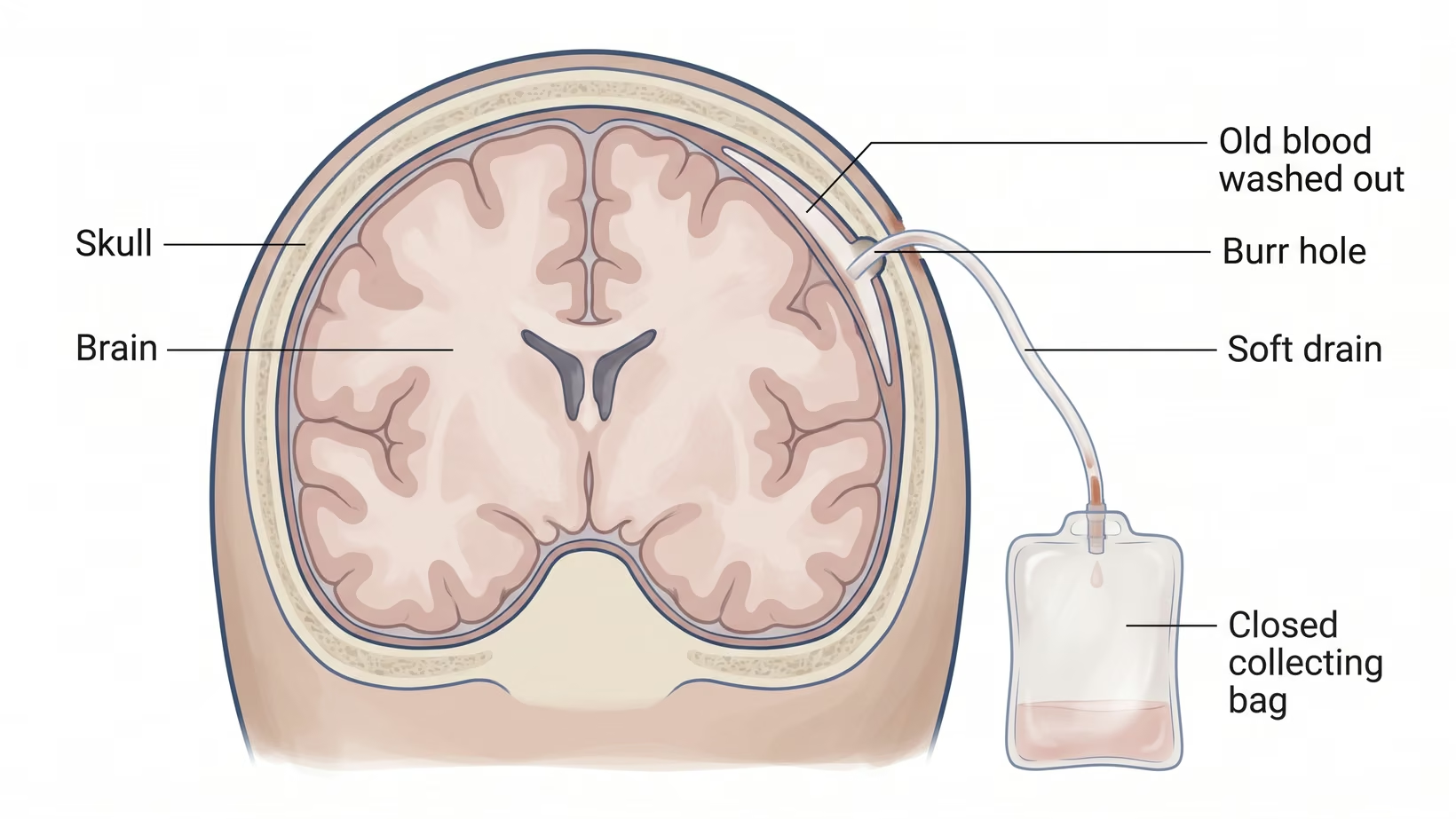
The standard operation for a chronic subdural hematoma that causes symptoms is burr-hole drainage with a closed drain. The surgeon makes a small hole in the skull and washes out the collection. A soft drain is left in the space for a day or two.
The drain is used because of one randomized trial. Repeat drainage was needed in 10 of 108 patients who had a drain, and in 26 of 107 who did not. Within six months, about 9 in 100 patients with a drain died (8.6%), against about 18 in 100 without one (18.1%).
In a Philippine series of 71 adults operated on at a tertiary medical center here, seven, about 10 in 100 (9.9%), had a recurrence.
The Steroid Question, Answered by a Trial
Families often ask about dexamethasone, a steroid. A large trial tested it in 748 patients with an average age of 74. At six months, about 84 in 100 of those given dexamethasone had a good outcome (83.9%), against about 90 in 100 of those given a placebo, a dummy pill (90.3%). The steroid group also had more harmful medical events during the trial.
Fewer patients on dexamethasone needed a repeat operation: about 2 in 100 (1.7%) against about 7 in 100 (7.1%). But overall, patients did worse on the drug. That is why it is not given routinely. If he has already been prescribed it, do not stop it on your own; ask the doctor who prescribed it how to stop it safely.
Blocking the Artery That Feeds It
A newer treatment, middle meningeal artery embolization, blocks the artery that supplies the membranes. A thin tube (catheter) is passed through the blood vessels to that artery, and the artery is then blocked. Two randomized trials support it when added to the standard treatment, but it is not available in most centers in the Philippines.
The Number Families Are Not Usually Told
A study that counted a whole population, not only surgical patients, found that about 9 in 100 (9.4%) died within 30 days and about 33 in 100 (32.9%) died within one year. In nearly half of the deaths in the first month, the collection was judged to be a partial or full cause. That is a reason to take falls, walking and safety at home seriously, not a reason to despair.
Bring and ask
What to Bring to the Consult, and What to Ask
Bring:
- The CT images on a disc or drive, not only the printed report
- A complete list of his medicines, including aspirin, clopidogrel, warfarin and the newer blood thinners, with the time of the last dose
- Recent blood test results if you have them
- A clear description of how he was three months ago: walking, talking, managing money, using the toilet
- The name and number of the doctor who usually prescribes his medicines
Ask:
- How thick is the collection, and is there shift across the midline
- Does he need an operation now, or can he be watched with a repeat scan
- If he is watched, when exactly is the next scan and who books it
- What happens to his blood thinner, and who decides when it restarts
- Will a drain be used
- How much will the admission cost, and what will PhilHealth cover here
- Which signs mean we come back before the next appointment
Frequently Asked Questions
Will My Father Need His Skull Opened?
Usually a chronic subdural hematoma does not need the skull opened. The standard operation is a burr hole, a small opening rather than a craniotomy (removal of a larger piece of bone), and the collection is washed out through it. A larger opening is used only for collections that cannot be cleared that way or that have come back.
Can It Just Go Away on Its Own?
A chronic subdural hematoma sometimes clears without surgery. Small collections in patients with few symptoms are watched rather than drained. But in the study described above, about 43 in 100 of those first managed without surgery later needed it. Watching is a valid plan only when the date of the next scan is set.
He Takes Aspirin. Do We Stop It?
Whether to stop aspirin is decided by the team treating him, and it is a hard decision. Do not stop it, skip a dose or change it on your own before you speak to them. Stopping blood thinners can cause complications from blood clots. Bring the exact name of the drug, the dose written on the box and the time of the last dose. Ask who will decide when it restarts.
How Long Before He Is Himself Again?
There is no reliable figure for how long recovery from a chronic subdural hematoma takes. No one can give you an exact number in advance. A more useful question is whether he is better this week than last week, and the family can answer that better than a scan can.
Can It Happen a Second Time?
Yes, a chronic subdural hematoma can come back. This is why the follow-up scan is booked before discharge. Keep that appointment even if he seems well. A collection can also form on the other side of the brain, because the shrinkage and stretched veins that allowed it are still there.
What to do now
What to Do Now
If your parent has a scan showing this and is awake, talking and stable, have the scan sent to a neurosurgeon today and see one in person within the next few days, not next month. If a warning sign appears before then, go to an emergency department now. You can book a consultation yourself, or his doctor can refer him.
- Bring the images and the other items in the checklist above
- Write down the blood thinner question and do not leave without an answer
- Set the date of the next scan before you go home
If he is drowsy, weak on one side, confused over hours, vomiting or having seizures, do not wait for a clinic appointment. Go to an emergency department now and bring the previous scan with you.
If there is no diagnosis yet and something about your father is not right some weeks after a bump on the head, see his doctor within the next day or two and ask whether a plain head CT is needed. If it is getting worse from day to day, or a warning sign appears, go to an emergency department now. It is a fast test, and this condition is usually easy to see once a scan is done.
This is not an illness to wait out at home. The most important thing now is for a doctor to see him while he can still talk clearly. You are right to be worried.
If you would like him to see me, my clinic for adults is at the Lung Center of the Philippines, Room 1101, Quezon Avenue, Quezon City, on Tuesday and Thursday from 2 PM. If the trip is too hard for him, we can talk in an online consult, booked on the consultation page.
See sources
- Pierre L, Julien C, Margetis K, Kondamudi NP. Subdural hematoma. StatPearls Publishing, updated 24 May 2026. https://www.ncbi.nlm.nih.gov/books/NBK532970/
- Edlmann E, Giorgi-Coll S, Whitfield PC, Carpenter KLH, Hutchinson PJ. Pathophysiology of chronic subdural haematoma: inflammation, angiogenesis and implications for pharmacotherapy. Journal of Neuroinflammation, 2017. PMID 28558815, DOI 10.1186/s12974-017-0881-y
- Santarius T, Kirkpatrick PJ, Ganesan D, et al. Use of drains versus no drains after burr-hole evacuation of chronic subdural haematoma: a randomised controlled trial. Lancet, 2009. PMID 19782872, DOI 10.1016/S0140-6736(09)61115-6
- Hutchinson PJ, Edlmann E, Bulters D, et al. Trial of dexamethasone for chronic subdural hematoma. New England Journal of Medicine, 2020. PMID 33326713, DOI 10.1056/NEJMoa2020473
- Davies JM, Knopman J, Mokin M, et al. Adjunctive middle meningeal artery embolization for subdural hematoma. New England Journal of Medicine, 2024. PMID 39565988, DOI 10.1056/NEJMoa2313472
- Fiorella D, Monteith SJ, Hanel R, et al. Embolization of the middle meningeal artery for chronic subdural hematoma. New England Journal of Medicine, 2024. PMID 39565980, DOI 10.1056/NEJMoa2409845
- Yang W, Huang J. Chronic subdural hematoma: epidemiology and natural history. Neurosurgery Clinics of North America, 2017. PMID 28325454, DOI 10.1016/j.nec.2016.11.002
- Hamou HA, Clusmann H, Schulz JB, Wiesmann M, Altiok E, Höllig A. Chronic subdural hematoma. Deutsches Arzteblatt International, 2022. PMID 35236548, DOI 10.3238/arztebl.m2022.0144
- Foppen M, Lodewijkx R, Slot M, Vandertop WP, Verbaan D. Chronic subdural hematoma with mild to moderate symptoms: the effect of initial treatment approach on clinical outcome. Brain and Spine, 2025. PMID 40115869, DOI 10.1016/j.bas.2025.104219
- Blaauw J, Meelis GA, Jacobs B, et al. Presenting symptoms and functional outcome of chronic subdural hematoma patients. Acta Neurologica Scandinavica, 2021. PMID 34448196, DOI 10.1111/ane.13518
- Kitya D, Punchak M, Abdelgadir J, Obiga O, Harborne D, Haglund MM. Causes, clinical presentation, management, and outcomes of chronic subdural hematoma at Mbarara Regional Referral Hospital. Neurosurgical Focus, 2018. PMID 30269586, DOI 10.3171/2018.7.FOCUS18253
- Mercado CMC, Maala JMD, Bayhon MV, et al. Predictors of recurrence of chronic subdural hematoma among adult Filipinos after surgery: developing a preliminary model for prognosis. Cureus, 2025. PMID 40395255, DOI 10.7759/cureus.82599
- Detran JEB, Villavicencio JB. Embolization of the middle meningeal artery as primary treatment for chronic subdural hematoma in older Filipino adults. Cureus, 2025. PMID 41439083, DOI 10.7759/cureus.97406
- Torio JER, Meceda EJA, Oanes FR, Cua MA, Elbangcol DR. Identification of middle meningeal artery confluence point in 3D CT scan images of Filipinos as pre-operative planning for management of chronic subdural hematoma. Journal of Cerebrovascular and Endovascular Neurosurgery, 2026. PMID 41593881, DOI 10.7461/jcen.2026.E2025.09.002
- Robinson D, Ding L, Stanton RJ, et al. Incidence and mortality of chronic subdural hematomas: a population-based study. Stroke, 2025. PMID 41287962, DOI 10.1161/STROKEAHA.125.053396