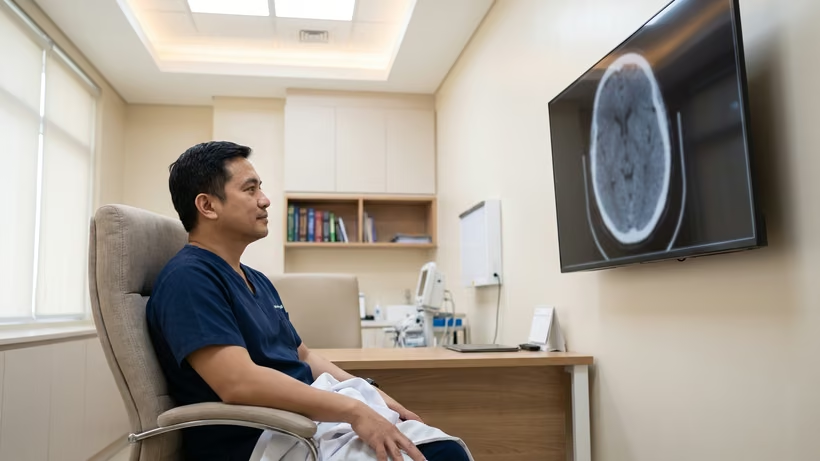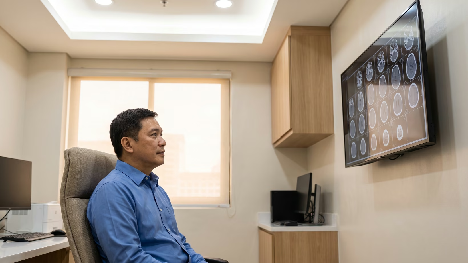

A normal cranial CT scan means the pictures showed no bleeding inside the skull, no swelling, no shift of the brain to one side, and no skull fracture at the moment the scan was taken. It does not mean no injury happened. A worsening headache, repeated vomiting, unusual sleepiness, confusion or weakness means going to the nearest emergency room now.
What is a cranial CT scan?
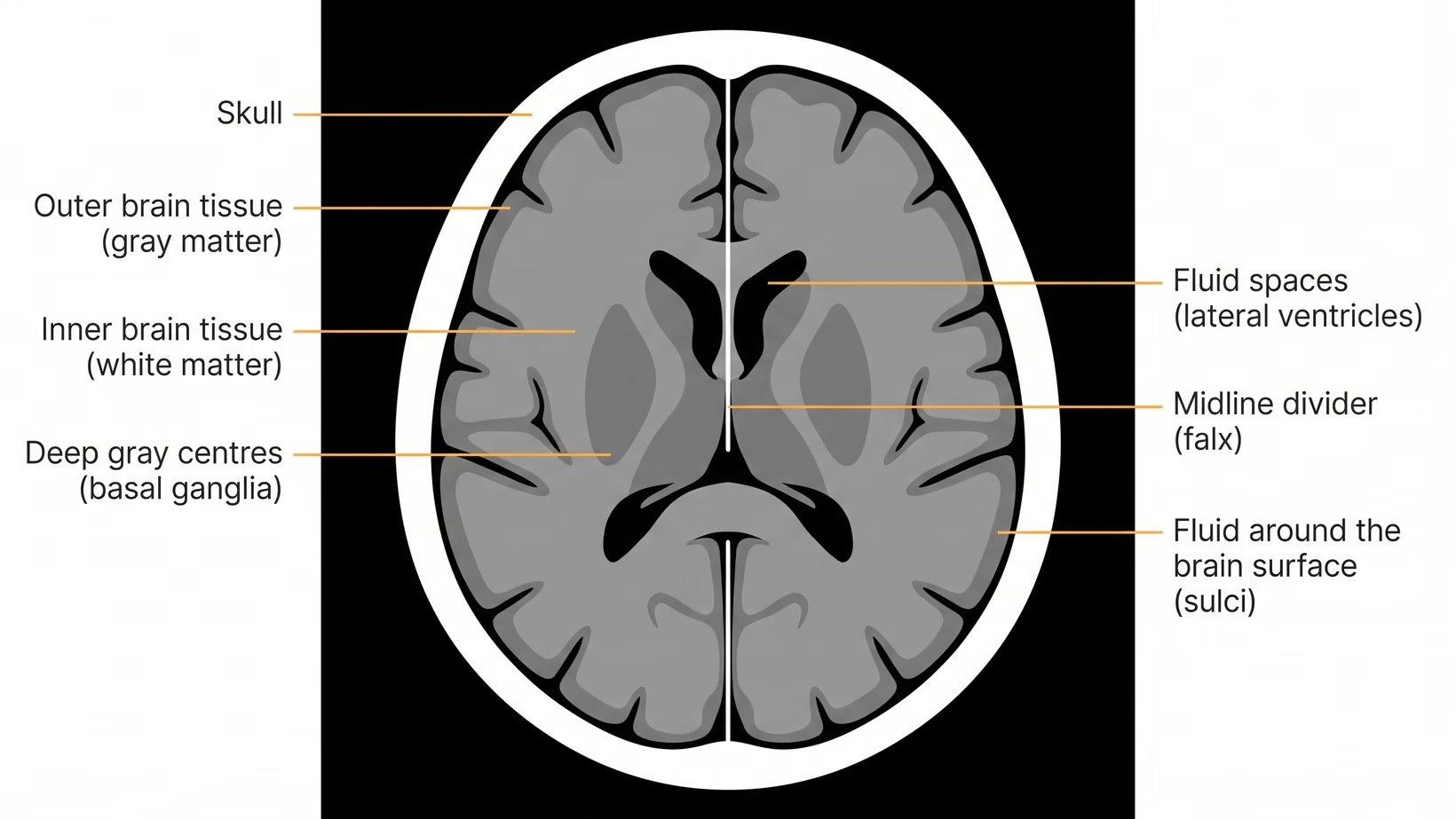
A cranial CT scan is a set of X-ray pictures, taken from many angles, which a computer builds into slices through the skull and the brain. It shows blood, bone, the fluid spaces inside and around the brain, and the shape of the brain tissue. It is the first scan ordered in an emergency room after a head injury.
It is not a test of how your brain is working. It is a picture of how your brain looks.
Cranial CT, head CT, CT brain: why one scan has several names
Cranial CT, head CT, CT scan of the head, CT brain and brain CT all name the same scan. The words that do change the scan are plain or with contrast. Plain means no dye was given, and that is the scan ordered after a head injury.
What does a normal cranial CT scan mean?
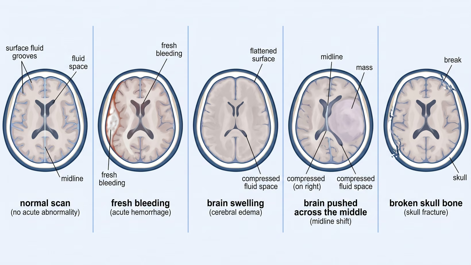
A normal result means the radiologist looked for fresh blood, swelling, a shift of the brain across the midline and a break in the skull bone, and found none of them. It is the common result after a head injury, not a lucky one. The radiologist is the doctor who reads the scan and writes the report.
In a Canadian study of 3,121 adults who were awake after a minor head injury, about 8 in 100 had an injury on the CT that mattered clinically, and about 1 in 100 needed urgent treatment for the brain injury, such as an operation. These were patients in Canadian emergency departments, so this is not a Philippine figure.
What a normal cranial CT scan does and does not rule out
A normal plain CT rules out the findings that would change what happens in the next few hours. How much it rules out depends on how soon it was done.
In adults with a sudden severe headache, a CT read by a qualified radiologist on a modern scanner within six hours of the headache starting found all 121 cases of subarachnoid hemorrhage, meaning bleeding into the fluid space around the brain. Across every patient in that study, whenever the scan was done, the CT found 92.9 in every 100 cases. Those patients had a sudden headache, not a head injury.
A concussion does not appear on a CT scan at all. It is diagnosed from your symptoms and the examination.
Sometimes a scan done for a head injury finds something unrelated to the injury. Many of these findings are watched with a planned repeat scan rather than treated. Ask your doctor which it is for you.
Seek care now
Go back to the emergency room even if the CT was normal
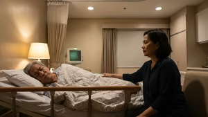
A normal scan describes one moment. A bleed can start or grow after the pictures were taken, so the scan does not end the need to watch the patient. Go to the nearest emergency room now, as soon as you notice any of the signs below, day or night. Do not wait for the morning. Bring the CT films and the report with you.
The warning signs to watch for after a normal scan
- A headache that keeps getting worse, or that wakes the patient from sleep
- Vomiting more than twice, or vomiting that starts hours after the injury
- Getting drowsy, hard to rouse, or harder to wake than before
- Confusion, not recognizing people, or speech that makes no sense
- Weakness, numbness or clumsiness on one side of the body
- A seizure, meaning a fit or convulsion
- Clear fluid or blood coming from the nose or the ear
- One pupil larger than the other, or new double vision
- New unsteadiness on standing or walking
- In a baby or small child: not feeding, inconsolable crying, or not behaving normally according to you
Bring and ask
Who to see, what to bring, and what to expect
Go to the nearest emergency room. If the hospital where the scan was done is about as close, go there, because that team already has the images and can compare a new scan with the first one.
Bring these:
- The CT films, and the imaging files on a CD or USB if the radiology department gave you one
- The printed radiology report
- A written list of all medicines, naming any blood thinner (anticoagulant) or aspirin-type medicine
- The date and time of the injury, and how it happened
- One relative who can stay as the bantay and answer questions
Expect a repeat examination first, not a repeat scan first. The doctor will check alertness, speech, strength and the pupils, and will decide from that whether a second scan or admission for observation is the safer answer. These checks look for a person who looked fine after a head injury and is now getting worse.
If there are no warning signs but the symptoms are not improving, see your doctor this week. You can book a consultation with a neurosurgeon, or your doctor can refer you. Bring the same films, report and medicine list.
If you would like me to see you, or your doctor wants to refer you to me:
- For an adult: my clinic at the Lung Center of the Philippines, Room 1101, Tuesday and Thursday from 2 PM.
- For a child: my clinic at the Philippine Children’s Medical Center (PCMC), Room 22, Wednesday from 2 PM.
- If the trip to Quezon City is not practical: an online consult, booked on the consultation page.
Plain or with contrast: which cranial CT scan was ordered?
A plain cranial CT is taken with nothing given to you. A cranial CT scan with contrast is taken after a dye is given, which makes blood vessels and certain growths brighter. After a head injury the plain scan is the one ordered, because blood, swelling, midline shift and skull fractures are all visible without any dye.
Why the request form says CT brain plain
The form says CT brain plain because the question is about the injury. A plain scan is also faster, needs no blood test first, and avoids a reaction to the dye.
Contrast is added when the question is about a growth, an infection or the blood vessels themselves. For fresh bleeding, swelling and fractures, the dye adds nothing and takes time to arrange.
What a plain CT scan of the brain can miss
A plain CT is good at showing blood and poor at showing a fresh blockage of a blood vessel. Pooled across small studies of patients scanned within 12 hours of stroke symptoms, a plain CT showed roughly 4 in 10 of the confirmed ischemic strokes, meaning strokes caused by a blocked vessel, while MRI showed nearly all of them. The studies were small, and the review itself calls the figures uncertain.
It also cannot show bleeding that started after the pictures were taken. For an early stroke, an MRI can answer what a CT cannot.
What does a CT scan of the brain actually show?
The scan separates tissues by how much X-ray each one absorbs. Bone appears white, fluid appears dark, and brain tissue sits between them in shades of gray. Fresh blood absorbs more X-ray than brain tissue, so it appears brighter than the brain around it, and the radiologist can see where it is and how much of it there is.
As blood gets older it breaks down and becomes less dense, so it appears gray, and then almost as dark as the fluid around the brain. The report calls recent blood acute, and it is the bright finding of a brain bleed after a head injury. It calls old blood chronic. Old blood can be missed by someone reading the images quickly, because its shade is close to the fluid beside it.
Reading a normal CT brain report, line by line
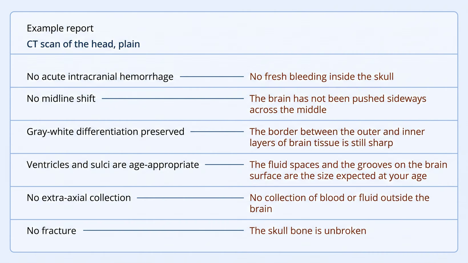
A normal cranial CT report is a list of structures with the word normal, or no abnormality, beside each one. Each line answers a plain question: is there fresh bleeding, has the brain been pushed sideways, is it swollen, are the fluid spaces the size expected at your age, and is the skull broken.
These phrases mean nothing is wrong:
- No acute intracranial hemorrhage: no fresh bleeding inside the skull. Intracranial means inside the skull.
- No midline shift: the brain has not been pushed sideways across the middle of the skull.
- Gray-white differentiation preserved: the border between the outer and inner layers of brain tissue is still sharp. That border blurs when the brain swells.
- Ventricles and sulci are age-appropriate: the fluid spaces inside the brain (ventricles) and the grooves on its surface (sulci) are the size expected at your age.
- No fracture: the skull bone is unbroken in the slices that were taken.
These phrases look alarming and are usually not:
- Atrophy: loss of brain volume. It is expected with age, and it is the reason the fluid spaces look wider on an older patient’s scan.
- Calcification: calcium in a structure that normally carries it, such as the pineal gland.
- Prominent ventricles: the fluid spaces are on the larger side. Whether that means anything depends on your age and your symptoms.
- Chronic ischemic change: old change in the small blood vessels of the brain. The radiologist would have written acute for anything recent.
None of these four lines describes an injury from today. Ask your doctor what each line of the brain scan report means for you.
No acute finding means nothing new or recent appears on the scan. Acute is a word about time, not about how serious something is. A report can say no acute finding on a brain with plenty of old change. It can also say it on a scan taken too early for a slow bleed to be visible.
The brain CT scan is normal but symptoms remain. What now?
A normal brain CT scan shows no bleeding, swelling, shift of the brain or skull fracture at the time it was taken. It cannot show a concussion, so symptoms can continue after a normal scan. Headache, dizziness, poor sleep and poor concentration after a head injury often come from the injury itself, not from something the scan missed.
A headache that keeps getting worse, or any other warning sign listed above, is different: go to the nearest emergency room now. A concussion is a common reason a person feels unwell with a normal scan.
Lying in a dark room until the symptoms disappear is not what is advised now. In a pooled analysis of seven studies of sport-related concussion, people given early physical activity or prescribed exercise recovered about four and a half days sooner than those who were not. Broken sleep went with slower recovery. Do not start hard exercise on your own. Ask your own doctor what is right for you before you change anything.
Symptoms that slowly improve week by week are a different situation from symptoms that do not change or that get worse. Go back to your doctor if the symptoms are not improving, if new symptoms appear, or if you cannot return to work or school. Any of the warning signs above means the emergency room instead.
What happens during a head CT scan?
You lie on your back on a flat table, with your head resting in a padded cradle. The table moves slowly into a short ring, the ring turns around your head, and the pictures are taken. Nothing touches you and nothing is injected for a plain scan. The one thing asked of you is to stay still.
You can breathe normally, and the ring is open at both ends, so you are not enclosed. Metal is removed first: earrings, hairpins, eyeglasses, hearing aids and removable dentures.
Bring a valid ID, the request form from the doctor, any PhilHealth or HMO card, and any previous films or discs. Before the scan, ask the billing section what the head CT scan costs at that hospital and what part your PhilHealth or HMO coverage pays. The price differs for a plain scan and a scan with contrast.
How long does a CT scan of the head take?
The scan of the head itself is short. Most of your time in the radiology department is registration, waiting for your turn, and being positioned on the table. The report takes longer than the scan, so in an emergency the doctor often reads the images on the screen first and the written report follows.
How much radiation is in a head CT scan, and is it safe?
A head CT uses X-rays, so it delivers a dose of radiation. The added risk from one scan is very small. The caution belongs mainly to children and to people having scan after scan, because the dose adds up across scans and the total matters more than any single one.
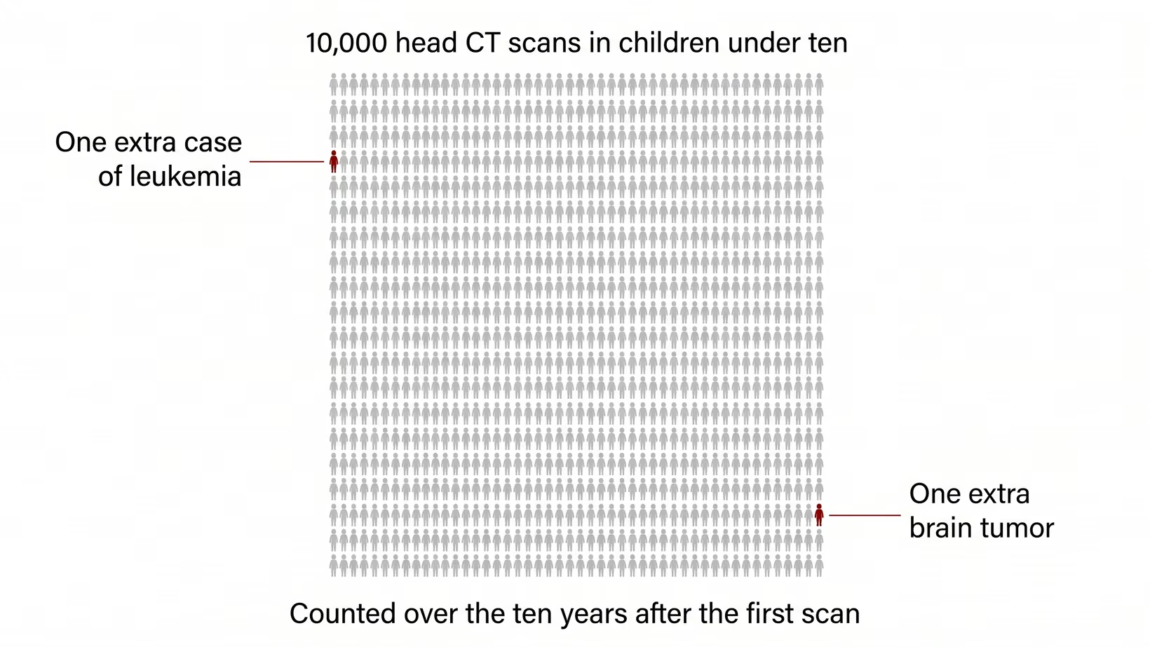
In a British study of children scanned between 1985 and 2002, about one extra case of leukemia and one extra brain tumor are expected for every 10,000 head CT scans in children under ten, counted over the ten years after the first scan.
This is why a doctor scans when the scan will change what happens next, and keeps the dose as low as possible. If the emergency doctor advises a scan, ask why; it is advised because the result will change what happens next. Tell the staff about every previous head scan, and tell them if you are pregnant or might be.
When does a head injury need a CT scan?
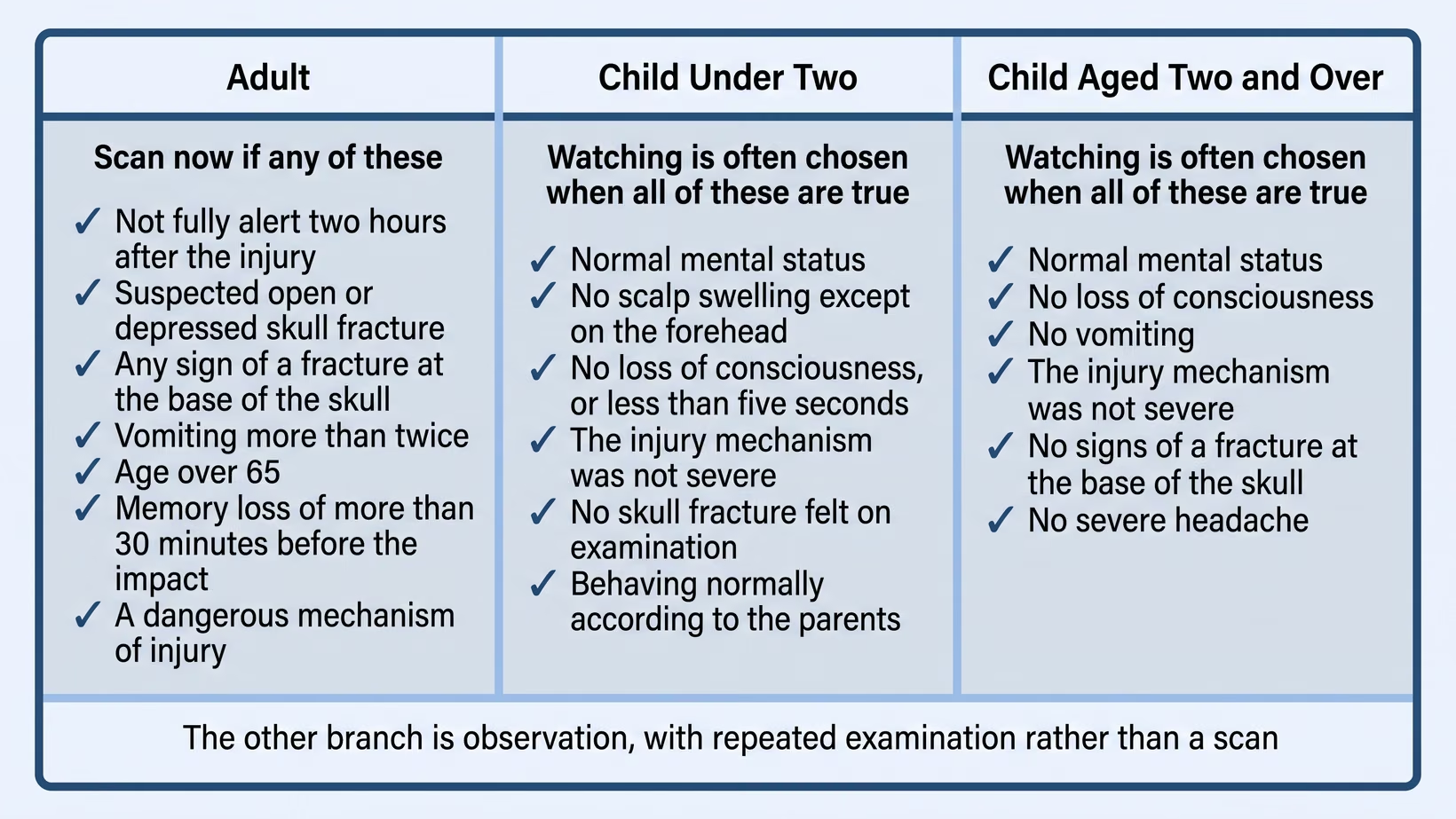
Not every head injury needs a CT scan. Emergency doctors use a short list of findings to decide, and a patient with none of them is often watched instead of scanned. For adults who are awake after a head injury, the list is the Canadian CT Head Rule. Children are checked with a different rule.
The rule was built from 3,121 adults in ten Canadian emergency departments. Five findings put an adult in the high-risk group: not back to full alertness two hours after the injury, a suspected open or depressed skull fracture, any sign of a fracture at the base of the skull, vomiting more than twice, and age over 65.
The five high-risk findings identified every patient in that study who needed urgent treatment for the brain injury. The true figure could be as low as 92 in 100. Having none of these findings does not make a bleed impossible.
Why a child with a minor head injury is often not scanned
An important brain injury is uncommon in a child who is alert after a head injury. In 42,412 children studied in North American emergency departments, 376 (about 9 in 1,000) had a brain injury that mattered clinically. Doctors use a separate rule to decide whether your child needs a CT scan after a head injury. None of this applies to a child who is drowsy, vomiting repeatedly, or hurt in a severe way. Take that child to the nearest emergency room now.
For referring physicians: what to send, and repeat scans
Send the time and mechanism of injury, the pupils, and the Glasgow Coma Scale (GCS, a bedside score of how the patient opens the eyes, speaks and moves) at the scene and now. Send the medicine list, naming any blood thinner (anticoagulant) or aspirin-type medicine (antiplatelet) and the last dose. Send the images, not only the report.
A repeat scan is for a patient who is changing, not for the calendar. Among 1,594 patients taking warfarin-type blood thinners who had a normal first head CT and were scanned again about 24 hours later, bleeding appeared on the second scan in about 6 in 1,000, and about 1 in 1,000 needed an operation or died. The authors leave out of that reassurance anyone with a serious mechanism of injury, heavy anticoagulation, antiplatelet medicine, or any worsening on examination.
For a patient with a normal first scan, repeated examination is what finds a new bleed, and a routine second scan 24 hours later is usually not needed. If a second scan has already been scheduled, keep it and ask the treating doctor before changing it.
Frequently asked questions
How long should I keep the CT films and the report?
Keep the CT films and the report permanently. Today’s scan is the comparison that saves a repeat scan and answers a question years from now, and the radiology department does not keep your copy for you. Ask for the files on a CD or USB, and bring them to every doctor who sees you, including the neurosurgeon if you are referred.
See sources
- Stiell IG, Wells GA, Vandemheen K, et al. The Canadian CT Head Rule for patients with minor head injury. Lancet. 2001. PMID 11356436.
- Kuppermann N, Holmes JF, Dayan PS, et al. Identification of children at very low risk of clinically-important brain injuries after head trauma: a prospective cohort study. Lancet. 2009. PMID 19758692.
- Perry JJ, Stiell IG, Sivilotti MLA, et al. Sensitivity of computed tomography performed within six hours of onset of headache for diagnosis of subarachnoid haemorrhage: prospective cohort study. BMJ. 2011. PMID 21768192.
- Pearce MS, Salotti JA, Little MP, et al. Radiation exposure from CT scans in childhood and subsequent risk of leukaemia and brain tumours: a retrospective cohort study. Lancet. 2012. PMID 22681860.
- Hauptmann M, Byrnes G, Cardis E, et al. Brain cancer after radiation exposure from CT examinations of children and young adults: results from the EPI-CT cohort study. Lancet Oncology. 2022. PMID 36493793.
- Brazzelli M, Sandercock PAG, Chappell FM, et al. Magnetic resonance imaging versus computed tomography for detection of acute vascular lesions in patients presenting with stroke symptoms. Cochrane Database of Systematic Reviews. 2009. PMID 19821415.
- Chauny JM, Marquis M, Bernard F, et al. Risk of delayed intracranial hemorrhage in anticoagulated patients with mild traumatic brain injury: systematic review and meta-analysis. Journal of Emergency Medicine. 2016. PMID 27473443.
- Leddy JJ, Burma JS, Toomey CM, et al. Rest and exercise early after sport-related concussion: a systematic review and meta-analysis. British Journal of Sports Medicine. 2023. PMID 37316185.