
A brain bleed after a head injury needs surgery when the blood is large enough to press on the brain, or when the person is getting worse. A neurosurgeon decides, from the CT scan and the patient together. Getting drowsier, weaker on one side, or harder to wake cannot wait: go to the emergency room now.
How big does a brain bleed have to be before surgery?
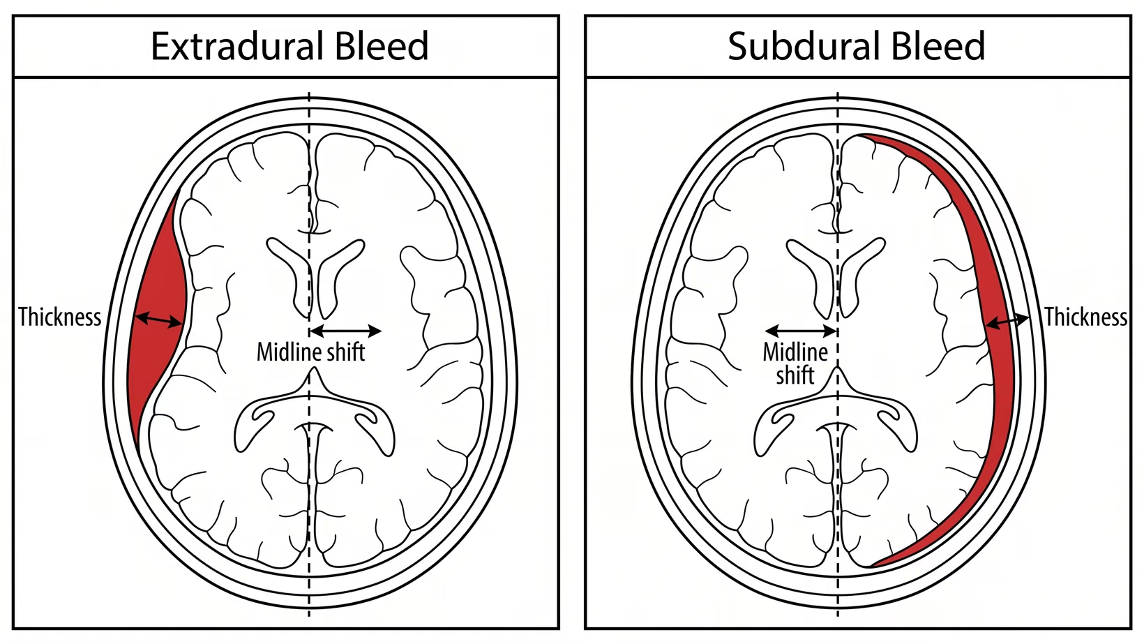
Each kind of brain bleed has its own cut-off for surgery. An extradural (epidural) hematoma, a bleed between the skull and the brain’s covering, is taken out above 30 cubic centimeters. An acute subdural hematoma, a bleed under that covering, is taken out above 10 mm thick or when it pushes the middle of the brain more than 5 mm to one side.
Both are taken out whatever the patient’s level of consciousness. The figures come from the surgical guidelines for traumatic brain injury.
The two numbers on the CT report: thickness and midline shift
Thickness is how deep the layer of blood is at its widest point, in millimeters. Midline shift is how far the structures that should sit in the middle of the head have been pushed to one side, also in millimeters.
Either number above its cut-off is a reason to operate on its own, without waiting for the patient to get worse. These numbers are recommendations about when to operate. They are not a measurement of how dangerous one particular bleed is.
Why the same size means different things by kind of bleed
An extradural bleed sits outside the dura, the tough covering of the brain. A subdural bleed spreads as a thin layer under the dura, over the surface of the brain.
A bruise or bleed inside the brain tissue, called a contusion, follows a third rule. There the decision rests more on the patient than on the picture. A size taken from one kind of bleed cannot be carried over to another kind of brain bleed after a head injury.
Why are they watching instead of operating?
They are watching because the bleed does not meet the cut-off for surgery, and watching is a treatment decision. For a bleed inside the brain tissue, a patient can be managed without surgery when the patient is not getting worse, the pressure inside the head is controlled, and the scan shows no significant pressing on the brain.
Watching does not mean going home. It means staying in hospital, with regular checks and a repeat CT scan to see whether the collection is growing. An operation on the head carries its own harm, so it is not done when the bleed does not need it.
Age, frailty, and what the person was like before the injury
Age and the state of the person before the injury are part of the decision, and sometimes the reason not to operate. An operation that a well 40-year-old recovers from can leave a frail 85-year-old alive but dependent on others for every part of care. Age by itself does not decide. Ask the surgeon what they expect for this patient.
That is why a surgeon asks what the person could do before the injury, and what the family would accept afterwards.
What happens if we say no to surgery?
If you say no to surgery, the team records the refusal and treats the patient with everything else it has: regular checks, control of the pressure inside the head where that is possible, medicines for pain and for seizures, and nursing care. Refusing is not permanent, and you can change your mind. If you do, tell the team at once.
The decision is looked at again whenever the patient changes or the family asks. Before you decide, ask the surgeon to show you the scan and to say what is expected without the operation, and ask about the options after refusing brain surgery.
Seek care now
Signs the bleed is getting worse: when to go to the ER now
Go to the nearest emergency room now, not in the morning, for any of these. The NICE head injury guideline is the source for advice to people sent home after a head injury, and your own doctor may add to this list:
- A headache that keeps getting worse
- Vomiting again and again
- Getting drowsier, or being hard to wake
- Confusion, or not knowing where they are
- Weakness in an arm, a leg, or one side of the face
- Slurred speech
- A seizure
- Clear fluid coming from the nose or the ear
If sent home: the signs that cannot wait until morning
A responsible adult should stay with someone sent home after a head injury. Do not let a person with a head injury sleep off the injury unwatched.
Bring the CT scan images and the report back with you, and the name of the hospital that did the scan, if they are at hand. Do not delay leaving to look for them. If the person takes a blood thinner, say so at the door of the emergency room.
How quickly does it have to happen?
Once there is a reason to operate, the guidelines say the operation is done as soon as possible. For taking out a clot, they set no number of hours, so there is no four-hour or six-hour rule to quote. The timing is as fast as the hospital can safely get the patient to the operating room.
A bleed in the posterior fossa, the part at the back and bottom of the skull, has no size cut-off. The guideline says to operate as soon as possible, because these patients can get worse quickly.
Who decides, the neurosurgeon or the ER doctor?
A neurosurgeon decides whether to operate. The emergency physician decides how fast the patient is examined, scanned, stabilized and referred. The emergency physician records the Glasgow Coma Scale score and the pupils, orders the CT scan, and manages the airway and the blood pressure.
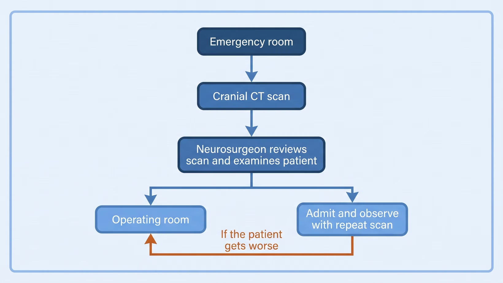
The neurosurgeon reads the scan images, examines the patient, and sets the plan: the operating room, or admission with a repeat scan. The guideline writers say their recommendations are not a complete protocol, and that judgment has to be added where the evidence is lacking.
Ask who the neurosurgeon on the case is, and ask that person for the plan in one sentence.
What happens when the hospital has no neurosurgeon on duty
Many hospitals in the Philippines have no neurosurgeon of their own. A survey published in 2023 counted 174 neurosurgeons in the country and found 35 provinces with none at all, which is 24 million people with no neurosurgeon in their province.
If you are in such a province, you can ask me for an online consult on the consultation page, and I can look at the scan with you.
The first hospital may do the CT scan and then telephone a neurosurgeon elsewhere. Ask whether a neurosurgeon has seen the actual images, and ask what the receiving hospital has said about the transfer to a hospital with a neurosurgeon.
What does consent for emergency brain surgery involve?
Consent for emergency brain surgery is a conversation and then a signature. The surgeon explains what the operation is meant to do, what it involves, what can go wrong, and what is expected without it. In an emergency that conversation is short, and it happens while the team is preparing.
Ask three things, in plain words: what the operation will do, what happens if it is not done, and who will be doing it. Signing the form does not end your right to ask.
Who can sign when the patient cannot
When the patient cannot understand, the closest available relative signs, usually a spouse, an adult child or a parent.
Put one decision-maker in front, because a surgeon cannot take a vote in a corridor. Bring any written instruction the patient left, or say what the patient had told the family.
Asking for a second opinion when there is no time
Asking for another neurosurgeon to look at the scan is reasonable. When the patient is getting worse there may be only minutes, so in practice it is the other neurosurgeon in the same hospital. Ask the first surgeon how long the decision can wait.
What head trauma surgery actually involves
Head trauma surgery takes the blood out and makes room for the brain. Under general anesthesia, the scalp is opened, an opening is made in the skull bone, and the blood is washed and suctioned out. Any bleeding point is stopped, and the skull and scalp are closed. Sometimes a drain is left in place.
Burr hole, craniotomy, and decompressive craniectomy
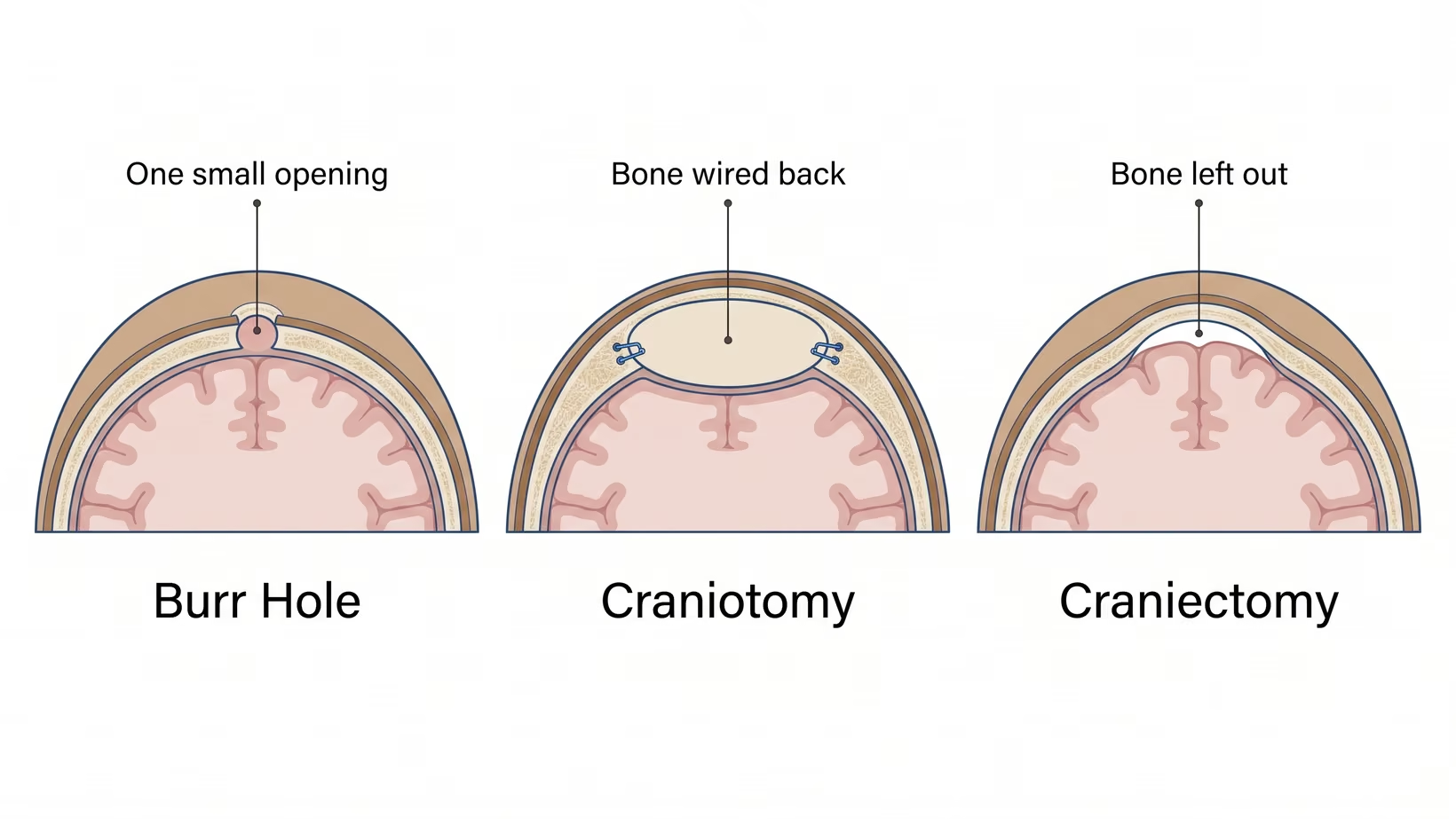
A burr hole is one small round opening in the skull bone, used to wash out thin blood. A craniotomy lifts a piece of skull bone out as a flap, so the whole collection and any bleeding point can be reached, and the bone is wired back at the end. A decompressive craniectomy leaves the bone out, so the brain has room to swell.
For an acute subdural bleed, the guideline recommends craniotomy, with or without removing the bone flap.
What the first days after the operation look like
Expect the first days in intensive care or close observation, with frequent checks and a repeat CT scan. A drain or a pressure monitor may be in place, and a machine may support breathing for a time.
How much the patient recovers is not known in the first days. The team watches whether the patient wakes, follows commands, and moves both sides.
Bring and ask
What emergency brain surgery costs and who can help pay
The cost depends on the operation, the hospital, the room, and the days in intensive care, so no web page can give you a figure. Ask the hospital directly, on the first day rather than at discharge. Ask these four:
- The billing section, for the bill so far and what is still expected
- The medical social service, for the hospital’s own assistance and the papers it needs
- The PhilHealth desk, for the case rate for this operation, and whether no balance billing applies
- The surgeon’s secretary, for the professional fees
Start the PhilHealth papers while the patient is still admitted.
Operative thresholds a referring doctor should know
The published cut-offs come from the Surgical Management of Traumatic Brain Injury guidelines. An extradural hematoma over 30 cubic centimeters, or an acute subdural hematoma over 10 mm thick or with more than 5 mm of midline shift, is evacuated whatever the Glasgow Coma Scale score. The cut-offs tell you when to refer and how urgently, not what the operation will achieve.
Extradural and acute subdural: the published cut-offs
A smaller extradural hematoma may be watched, with serial CT scans and close neurological observation in a center that has a neurosurgeon. The conditions are thickness under 15 mm, midline shift under 5 mm, a score above 8, and no focal deficit, meaning no weakness traceable to one part of the brain. A patient in coma, meaning a score under 9, with an extradural bleed and anisocoria, meaning pupils of unequal size, is evacuated as soon as possible.
A comatose patient with an acute subdural hematoma that is thinner and less shift is still operated on if any of these is present:
- The score fell by two or more points between the injury and admission
- The pupils are asymmetric, or fixed and dilated
- The intracranial pressure, meaning the pressure inside the skull, is above 20 mm Hg
The guideline recommends intracranial pressure monitoring for every comatose patient with an acute subdural bleed.
Contusions, posterior fossa lesions, and depressed fractures
A parenchymal lesion, meaning an area of damage inside the brain tissue such as a bruise or bleed, is operated on for deterioration caused by it, for raised pressure that does not respond to medical treatment, or for mass effect on CT, meaning the lesion pressing on the brain around it.
For a frontal or temporal contusion, the figure is a volume over 20 cubic centimeters, in a patient with a score of 6 to 8 and at least 5 mm of shift or squeezing of the cisterns. Any lesion over 50 cubic centimeters is also operated on.
A posterior fossa lesion is operated on for mass effect on CT or for deterioration traceable to the lesion. Mass effect here means distortion or loss of the fourth ventricle, one of the fluid spaces inside the brain, compression of the basal cisterns, or obstructive hydrocephalus, meaning fluid blocked and backing up inside the brain.
An open depressed skull fracture, depressed deeper than the thickness of the skull, is operated on early to prevent infection. A closed depressed fracture may be managed without surgery.
Anticoagulants and antiplatelets change the decision
A blood thinner changes what has to be arranged before an operation: an anticoagulant slows clotting, and an antiplatelet stops platelets from sticking together. Have ready the name of the medicine, the dose schedule, and the time of the last dose, and say it on the referral call.
What to send with the patient, and what to say on the phone
On the phone, give the mechanism and time of the injury, the score now and whether it has fallen, the pupils, and what the CT shows. In a patient in coma, a falling score or unequal pupils can each be a reason to operate.
Send with the patient:
- The actual CT images, on a disc or a USB, not only the printed report
- The time of the injury and the time of the scan
- The score at first contact and the score now, with the times
- Pupil size and reaction on both sides
- The list of medicines, with any anticoagulant or antiplatelet named, and the time of the last dose
- Airway status, blood pressure, and oxygen saturation
- A contact number for the relative who can consent
What to do now
What to do now
If the person has any of the warning signs above, go to the emergency room now, not in the morning.
If a bleed was found and the person is at home with none of the warning signs, ask today for a neurosurgeon to look at the scan: book a consultation, or ask your doctor to refer you. Bring the actual CT images and report, the name of the hospital that did the scan, and the list of medicines with any blood thinner named, so the first neurosurgery appointment can start from them.
You may also see me for this. I see adults at the Lung Center of the Philippines in Quezon City on Tuesday and Thursday from 2 PM. I see children at the Philippine Children’s Medical Center on Wednesday from 2 PM.
If you live too far to come to the clinic, you can book an online consult with me on the consultation page.
Frequently asked questions
How long does a brain bleed operation take?
How long a brain bleed operation takes depends on which operation is done and on what is found inside. Ask the surgeon for an estimate before the patient goes in, and ask where the family should wait.
Can a brain bleed go away without surgery?
A brain bleed below the cut-offs for surgery can be managed without an operation, with intensive monitoring and repeat scans in hospital. That is not the same as going away by itself. The guidelines describe watching the patient in hospital, not sending them home to see what happens.
Is surgery for a brain bleed safe for an elderly patient?
Age by itself does not decide whether surgery for a brain bleed is right for an elderly patient. The surgeon weighs what the person could do before the injury, their other illnesses, and what the family would accept as a result. Ask the surgeon what they expect for this patient.
What is the biggest brain bleed that can be watched?
The biggest brain bleed that can be watched differs with the kind of bleed. An acute subdural bleed above 10 mm thick or with more than 5 mm of shift is a reason to operate. An extradural bleed above 30 cubic centimeters is a reason to operate.
Will the skull bone be put back?
The skull bone is put back in a craniotomy, wired in place at the end of the same operation. It is left out in a decompressive craniectomy, so that the swollen brain has room. The surgeon makes that choice during the operation.
How soon will we know if the surgery worked?
The first sign that the surgery worked is the repeat CT scan, and whether the patient wakes and follows commands. How much function returns is known over weeks rather than in the recovery room.
See sources
- Bullock MR et al, for the Surgical Management of Traumatic Brain Injury Author Group. Surgical management of acute epidural hematomas. Neurosurgery 2006;58(3 Suppl):S7-15. PMID 16710967.
- Bullock MR et al, for the Surgical Management of Traumatic Brain Injury Author Group. Surgical management of acute subdural hematomas. Neurosurgery 2006;58(3 Suppl):S16-24. PMID 16710968.
- Bullock MR et al, for the Surgical Management of Traumatic Brain Injury Author Group. Surgical management of traumatic parenchymal lesions. Neurosurgery 2006;58(3 Suppl):S25-46. PMID 16540746.
- Bullock MR et al, for the Surgical Management of Traumatic Brain Injury Author Group. Surgical management of depressed cranial fractures. Neurosurgery 2006;58(3 Suppl):S56-60. PMID 16540744.
- Bullock MR et al, for the Surgical Management of Traumatic Brain Injury Author Group. Surgical management of posterior fossa mass lesions. Neurosurgery 2006;58(3 Suppl):S47-55. PMID 16540745.
- Carney N, Totten AM, O’Reilly C et al, Brain Trauma Foundation. Guidelines for the Management of Severe Traumatic Brain Injury, Fourth Edition. Neurosurgery 2017;80(1):6-15. PMID 27654000.
- Hutchinson PJ et al, RESCUEicp Trial Collaborators. Trial of decompressive craniectomy for traumatic intracranial hypertension. New England Journal of Medicine 2016;375(12):1119-30. PMID 27602507.
- Cooper DJ, Rosenfeld JV, Murray L et al, DECRA Trial Investigators. Decompressive craniectomy in diffuse traumatic brain injury. New England Journal of Medicine 2011;364(16):1493-502. PMID 21434843.
- Hutchinson PJ et al, RESCUE-ASDH Trial Collaborators. Decompressive craniectomy versus craniotomy for acute subdural hematoma. New England Journal of Medicine 2023;388(24):2219-2229. PMID 37092792.
- Baticulon RE, Lucena LLN, Gimenez MLA, Sabalza MN, Soriano JA. The neurosurgical workforce of the Philippines. Neurosurgery 2023;94(1):202-211. PMID 37931081.
- National Institute for Health and Care Excellence. Head injury: assessment and early management. NICE guideline NG232, 2023. https://www.nice.org.uk/guidance/ng232