

A brain bleed after a head injury is one of four types: outside the brain’s tough covering (epidural), under it (subdural), in the fluid over the brain (subarachnoid), or inside the brain. Which is worse depends on how fast the blood collects and how hard it presses. Getting harder to wake or weak on one side means the ER now.
What are the four types of brain bleed a head CT can show?
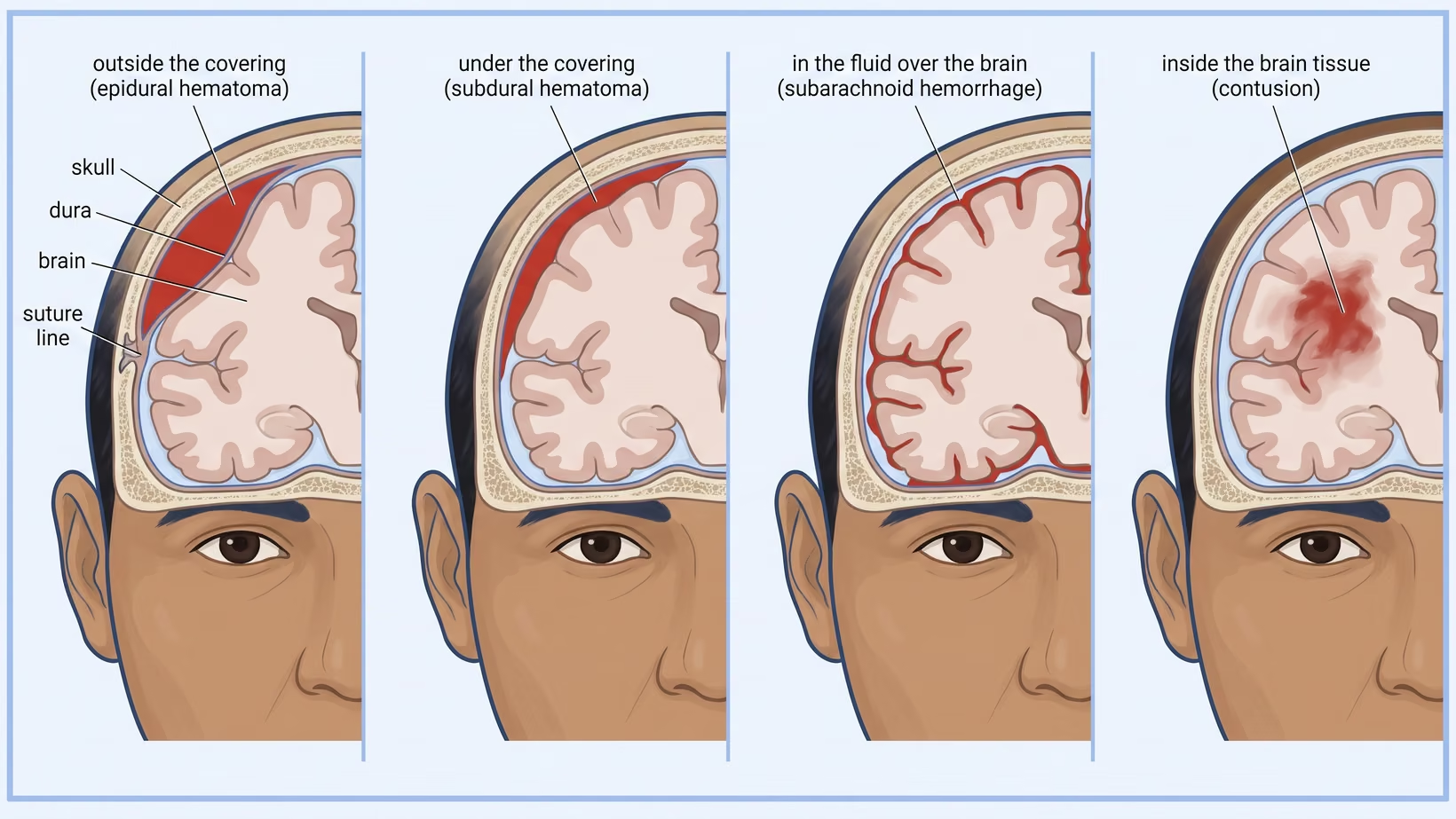
A head CT can show four bleeds, named for the space they fill. An epidural bleed is outside the dura, the tough membrane lining the skull like a leather bag. A subdural bleed is between the dura and the brain. A subarachnoid bleed is in the fluid over the brain. A contusion is inside the brain tissue.
A contusion is also written as an intracerebral bleed. Blood can also collect in the ventricles, the fluid chambers deep inside the brain, and a report may name that as well.
The name tells you where the blood is. It does not by itself tell you how serious this is.
Which bleed is on my CT report?
Your CT report names the bleed by its medical term, and each term below has its plain meaning. Hematoma means a collection of clotted blood. Hemorrhage means bleeding. These are the words a Philippine CT report uses, and knowing them is the first step in how to read a cranial CT result.
- Extradural hematoma, epidural hematoma, or EDH: blood outside the dura.
- Subdural hematoma, or SDH: blood between the dura and the surface of the brain.
- Subarachnoid hemorrhage, or SAH: blood in the fluid spaces over the brain.
- Intraparenchymal hemorrhage, intracerebral hemorrhage, contusion, hemorrhagic contusion: blood inside the brain tissue.
- Subgaleal hematoma, and cephalohematoma in a newborn: blood in the scalp, outside the skull bone. These are not a bleed inside the skull at all.
A report may also name the side and the lobe, the thickness in millimeters, and the midline shift, which is how far the middle of the brain has been pushed across. Those three are the numbers a surgeon reads first.
Seek care now
When to go to the emergency room right away
Go to the emergency room now, not in the morning, whether or not the person has already been seen, if they show any of the signs below. They apply whether you are watching a baby, an adult after a motorcycle crash, or an elderly parent after a fall. Go to the nearest emergency room that has a CT scanner.
- Getting harder to wake, or not fully awake between checks.
- Vomiting again and again.
- A seizure after the injury.
- New weakness, numbness or clumsiness on one side, or slurred speech.
- Pupils of different sizes, or one eye no longer moving with the other.
- A headache that keeps getting worse.
- Confusion, strange behavior, or not knowing where they are.
- Clear fluid or blood from the nose or ear, bruising behind an ear, or bruising around both eyes.
- In a baby: a soft spot on the head that is tense or bulging while the baby is calm, or feeding much less than usual.
Do not delay leaving to look for the papers. If they are at hand, bring the films or the disc from any earlier scan, the printed report, and a written list of every medicine the patient takes, blood thinners included. Photograph the report with your phone before you leave any hospital. On arrival, expect the level of consciousness to be scored, the pupils checked, and a CT scan of the head.
If a hilot is the first stop, the hours spent there are hours not spent on a scan. A hilot cannot find a bleed inside the skull. Go to the hospital first, and nobody needs to be blamed for not having known that.
Is a subdural or an epidural bleed worse?
Neither an epidural nor a subdural bleed is always worse. The two are measured differently. An acute subdural bleed thicker than 10 mm, or pushing the midline over by more than 5 mm, is removed in the operating room. An epidural bleed is removed when its volume is over 30 cubic centimeters. Both rules apply whatever the level of consciousness.
Only the thickness can be compared between the two. A subdural bleed is removed at a thickness above 10 mm. An epidural under 15 mm thick, with under 30 cubic centimeters and under 5 mm of shift, may be watched in an awake patient with no weakness on one side. Watching means repeat scans and close observation in a center that has a neurosurgeon. The epidural removal rule is a volume over 30 cubic centimeters.
So a subdural is operated on at a smaller thickness.
Why an easier-to-remove bleed can be more urgent
Urgency is set by the patient, not by the name of the bleed. An epidural bleed in someone who has become unconscious, and whose pupils have become unequal in size (anisocoria), is removed as soon as possible. A craniotomy, an operation that lifts a window of skull bone, removes the clot more completely.
Why does a subdural bleed come on slowly?
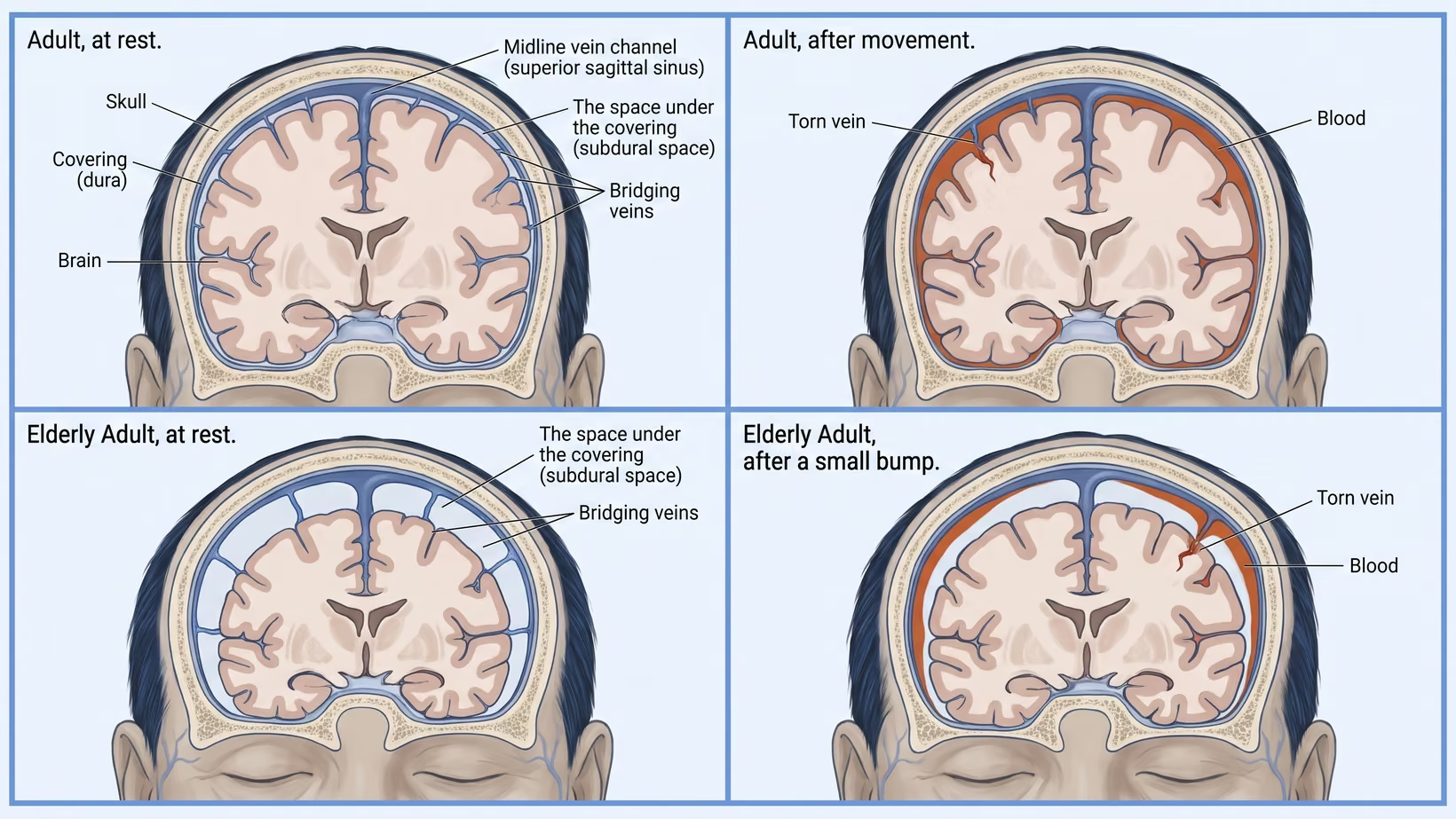
A subdural bleed can come on slowly, and the collection can take weeks to show.
The slow collection is called a chronic subdural hematoma. If an older person becomes more confused, drowsy or weak on one side, or has a headache that keeps getting worse, in the weeks after a fall, even a small one, go to the emergency room now.
Why the shape of the bleed on the scan names it
The shape of a bleed follows the space it fills. An epidural bleed usually looks like a lens on the scan. A subdural bleed usually looks like a thin crescent along the curve of the brain.
The shape is a strong clue and not a diagnosis on its own. The radiologist reads it together with where the bleed is and what it is pressing on.
Why one fall can cause two different bleeds
One fall can cause two different bleeds because one impact can injure two places at once. The blow can tear a vessel at the point of contact. The same movement can throw the brain against the inside of the skull on the opposite side, bruising it there.
One scan can then show a bleed under the point of impact and a contusion on the far side. Doctors call that pattern coup and contrecoup, the French words for the blow and the counter blow. A report naming two findings does not mean there were two accidents.
How a brain bleed changes over the first day
A brain bleed can grow over the first day, so the first scan describes the patient only at that point in time. The decision to operate follows the course over the hours after it. That is why the examination is repeated and the scan may be repeated, and why a small bleed that is not removed is still watched closely.
In someone in a coma with an acute subdural bleed under the size threshold, the guideline still says to operate if the person has deteriorated: the coma score fell by two or more points between the injury and arrival at hospital, the pupils are unequal or wide and unreactive, or the pressure inside the head goes above 20 mm Hg. Anyone in a coma with an acute subdural bleed has the pressure inside the head monitored, and when surgery is needed it is done as soon as possible.
What it means if you were sent home with a small bleed
In the guidelines, a bleed that is not operated on is still a bleed that is watched with repeat scans in hospital. Being sent home with a small brain bleed is a separate decision, made by the team who examined the patient. It comes with instructions about who watches and when to come back. Follow those instructions exactly. If any of the warning signs above appear before then, go to the emergency room now, without waiting for the planned visit.
Does a blood thinner change which bleed you get?
It changes how carefully you are checked after a bump on the head. The Scandinavian guideline for adults with a mild or moderate head injury makes being on an anticoagulant, a blood thinner that slows clotting, a reason for a CT scan on its own. If you take a blood thinner and hit your head, go to an emergency room that has a CT scanner today to be checked, even if you feel well.
A normal first scan is not the end of it for everyone on a blood thinner. In 18 pooled hospital studies of 4,613 adults who had a mild head injury and a normal first scan, a bleed appeared on a repeat scan in about two of every 100. Of those 4,613 patients, 67 had such a delayed bleed, 11 needed an operation and six died.
Whether your scan is repeated is a decision for the doctor who saw you. Stopping or restarting a blood thinner is that team’s decision too, and not one to make at home.
Which bleeds a provincial hospital must transfer
Any bleed that meets an operating threshold has to reach a neurosurgeon, and in much of the Philippines that means a transfer. A national survey counted 174 neurosurgeons for the whole country, about one for every 600,000 people. Thirty-five provinces had none at all and nine had one only part of the time.
That left about 24 million people with no local access. A small epidural bleed may be watched only with repeat CT scans and close neurological observation in a center that has a neurosurgeon. A hospital without one cannot offer that, so the same scan that would be watched in Manila is a transfer in a district hospital.
Send the images with the patient, on the disc and not only as a printed report. Write down the coma score at the scene, the blood pressure, and the full list of medicines including blood thinners. The first sentence to the receiving neurosurgeon carries the age, the time of injury, the current coma score, the pupils, which bleed it is with its thickness or volume, and the midline shift in millimeters.
Bring and ask
What to ask about cost before a transfer
Ask before the ambulance leaves, and ask the person whose job it is to answer. No two hospitals charge the same.
- Ask the attending doctor what operation is likely to be needed.
- Ask the admitting officer of the receiving hospital what deposit is required, and whether the transfer can proceed while that is being arranged.
- Ask the medical social worker what assistance the receiving hospital gives, and which papers you have to bring to apply for it.
- Ask the PhilHealth desk which benefits apply to this admission and this operation, and what documents each one needs.
- Ask for an itemized bill at each hospital, and ask who pays for the ambulance.
Surgical thresholds by bleed type
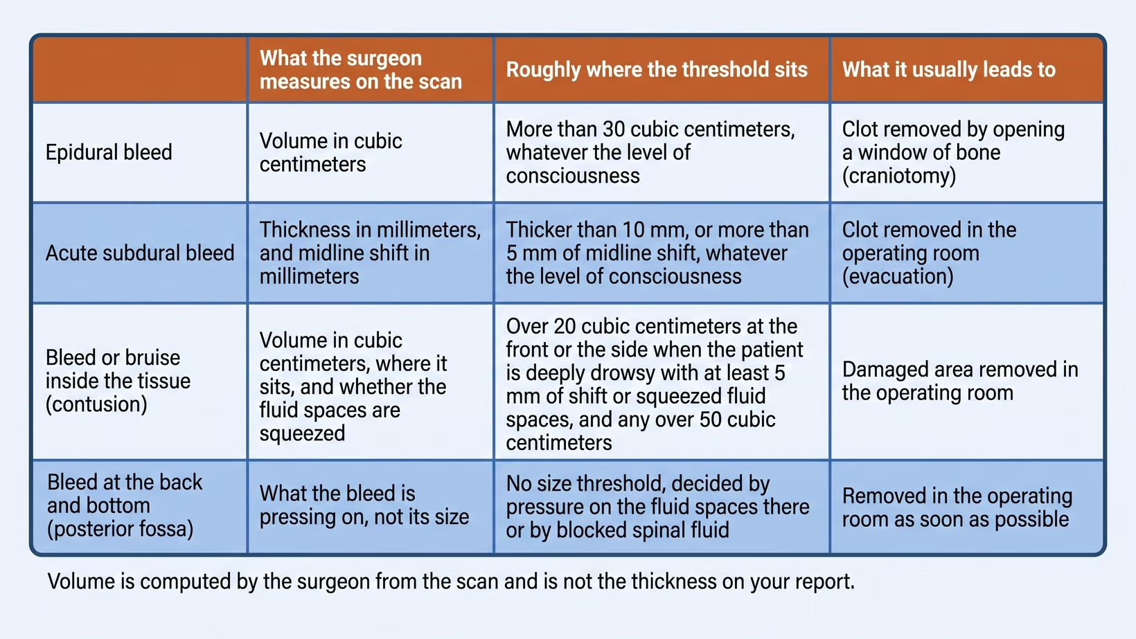
Each type of bleed has its own measurement and its own trigger to operate. A threshold is a trigger to operate. It does not mark a line between safe and dangerous. The surgeon computes volume from the scan, so the thickness in millimeters on your report is a different number.
- Epidural bleed: more than 30 cubic centimeters is removed whatever the level of consciousness.
- A smaller one, under 15 mm thick with under 5 mm of shift, in an awake patient with no weakness on one side, may be watched with repeat scans in a center that has a neurosurgeon.
- In coma with unequal pupils it is removed as soon as possible.
- Acute subdural bleed: more than 10 mm thick, or more than 5 mm of midline shift, is removed whatever the level of consciousness.
- Acute subdural bleed in a patient in a coma, meaning a score below 9 on the Glasgow Coma Scale (the 15-point bedside score of alertness), a smaller one is removed if the score fell by two or more points between the injury and arrival at hospital,
- if the pupils are unequal, or wide and unreactive, or if the measured pressure inside the head goes above 20 mm Hg.
- That pressure comes from a monitor placed in the skull in an intensive care unit, so nobody can know it at home.
- Bleed or bruise inside the brain tissue: it is operated on if the patient is getting steadily worse because of it, if the pressure inside the head cannot be controlled with medicines, or if the scan shows it pressing on the brain.
- A frontal or temporal contusion over 20 cubic centimeters is operated on in a patient in a coma (coma score 6 to 8) when the scan also shows at least 5 mm of shift or squeezed cisterns, and any over 50 cubic centimeters is operated on.
- Only when none of these are present is it watched with intensive monitoring and repeat scans.
- Bleed in the posterior fossa, the compartment at the back and bottom of the skull that holds the brainstem and the cerebellum: no size threshold.
- It is operated on when the fourth ventricle, a fluid space between the brainstem and the cerebellum, is distorted or pushed out of place; when the basal cisterns, the fluid spaces around the brainstem, are squeezed or no longer visible; or when spinal fluid is blocked from draining.
- It is then operated on as soon as possible, because these patients can get worse quickly.
What to do now
What to do now
If the person has any of the warning signs above, go to the emergency room now, not in the morning.
If a bleed was found and you want a neurosurgeon to look at it, you can book a consultation, or your doctor can refer you. See a neurosurgeon and bring the films or the disc, the printed report, the name of the hospital that did the scan, and a written list of every medicine the patient takes, blood thinners included.
Frequently asked questions
Which brain bleed is the most dangerous after a head injury?
No one brain bleed is the most dangerous after a head injury, and the guidelines do not rank them by deaths. Measured by thickness, an acute subdural is operated on at a smaller size than an epidural. A bleed in the posterior fossa is judged by what it presses on rather than by size.
Can a brain bleed stop on its own without surgery?
Many brain bleeds are never operated on, and the guidelines describe watching rather than operating for bleeds below the thresholds. That watching means repeat CT scans and close observation in hospital, not healing at home. A small epidural bleed may be watched this way only in a center that has a neurosurgeon.
How long after hitting your head can a bleed still appear?
A brain bleed can appear on a repeat scan after hitting your head even when the first scan was normal. In pooled studies of adults on blood thinners after a mild injury, that happened in about two of every 100. A slow subdural collection in an older person can be found weeks after a bump that nobody remembers. New confusion, drowsiness, weakness on one side or a worsening headache in an older person means the emergency room now, even if no fall is remembered.
Does a brain bleed always need an operation?
A brain bleed does not always need an operation. The measurement on the scan decides, and so does the patient’s course. In someone who is in a coma, even a small subdural bleed is removed if the coma score fell by two or more points between the injury and arrival at hospital, the pupils are unequal or wide and unreactive, or the pressure inside the head rises above 20 mm Hg.
Do children get the same kind of bleed as adults?
A head injury in a child can cause the same four bleeds as in an adult, because a child’s skull holds the same four spaces. The guidelines quoted on this page were written for adults, so none of the numbers above should be read as a child’s number.
See sources
- Bullock MR, Chesnut R, Ghajar J, et al, for the Brain Trauma Foundation and the AANS/CNS Section on Neurotrauma. Surgical management of acute epidural hematomas. Neurosurgery 2006;58(3 Suppl):S7-15. PMID 16710967
- Bullock MR, Chesnut R, Ghajar J, et al. Surgical management of acute subdural hematomas. Neurosurgery 2006;58(3 Suppl):S16-24. PMID 16710968
- Bullock MR, Chesnut R, Ghajar J, et al. Surgical management of traumatic parenchymal lesions. Neurosurgery 2006;58(3 Suppl):S25-46. doi:10.1227/01.NEU.0000210365.36914.E3
- Bullock MR, Chesnut R, Ghajar J, et al. Surgical management of posterior fossa mass lesions. Neurosurgery 2006;58(3 Suppl):S47-55. doi:10.1227/01.NEU.0000210366.36914.38
- CRASH-3 trial collaborators. Effects of tranexamic acid on death, disability, vascular occlusive events and other morbidities in patients with acute traumatic brain injury (CRASH-3): a randomised, placebo-controlled trial. Lancet 2019. doi:10.1016/S0140-6736(19)32233-0
- Hutchinson PJ, Kolias AG, Timofeev IS, et al. Trial of decompressive craniectomy for traumatic intracranial hypertension (RESCUEicp). New England Journal of Medicine 2016. doi:10.1056/NEJMoa1605215
- Hutchinson PJ, Edlmann E, Bulters D, et al. Trial of dexamethasone for chronic subdural hematoma (Dex-CSDH). New England Journal of Medicine 2020. doi:10.1056/NEJMoa2020473
- Unden J, Ingebrigtsen T, Romner B, for the Scandinavian Neurotrauma Committee. Scandinavian guidelines for initial management of minimal, mild and moderate head injuries in adults: an evidence and consensus-based update. BMC Medicine 2013. doi:10.1186/1741-7015-11-50
- Baticulon RE, et al. The neurosurgical workforce of the Philippines. Neurosurgery 2023. doi:10.1227/neu.0000000000002630
- El Hadwe S, Assamadi M, Barrit S, et al. Delayed intracranial hemorrhage of patients with mild traumatic brain injury under antithrombotics on routine repeat CT scan: a systematic review and meta-analysis. Brain Injury 2022. doi:10.1080/02699052.2022.2065034