

A chronic subdural hematoma, blood that has collected slowly under the lining of the skull, can be absorbed without an operation. Watching it with repeat scans is a treatment choice. It works best when the blood layer is thin, the brain is not pushed aside, and symptoms are mild. New drowsiness, confusion or weakness means the nearest emergency room now.
Can a Subdural Hematoma Absorb on Its Own?
Yes, a subdural hematoma can absorb on its own, and many chronic ones do. Guideline reviews put the share that clears without treatment at about 4 in 10 (roughly 40 percent). Among people with no symptoms or only mild ones, the reported range runs from about 2 in 10 to about 5 in 10.
Ang hirap pong pakinggan ng “babantayan muna natin” kapag ang naririnig ninyo ay dugo sa ulo. Pero ang pagbabantay po ay may plano, may petsa, at may listahan ng senyales na hindi hinihintay.
What Your Body Is Doing With the Blood
A subdural hematoma is blood collecting between the dura, the tough lining inside the skull, and the thin layer over the brain. In a chronic collection the body is already breaking that blood down. At the same time the membrane around it grows fragile new vessels that keep refilling it.
Watching works only if the body clears the blood faster than the new vessels refill it. The repeat scan checks which one is happening.
Why an Acute Bleed Is a Different Question
Timing changes the answer. Blood seen within 72 hours of an injury is acute, a collection found after about three weeks is chronic, and the span between is subacute.
An acute bleed can expand fast, so there the decision is made in hours. The chronic collection has had weeks to settle, and it is the one most often watched.
Seek care now
Which Signs Mean the Emergency Room Now?
These signs mean a subdural hematoma that is being watched cannot wait for the next appointment. Waiting at home still carries a risk, and it is lower only while someone who lives with the patient watches for these signs every day. If any one appears, go to the nearest emergency room now and bring the scan. If one with a neurosurgeon on call is just as close, choose it, but do not drive past an emergency room to reach it.
- New or worsening drowsiness, or someone hard to rouse
- Confusion or a personality change that is new today
- Weakness, numbness or clumsiness on one side
- Slurred speech, or trouble finding words
- A headache suddenly much worse, or one that wakes the person from sleep
- Repeated vomiting
- A seizure of any kind
- A new fall, or any fresh bump to the head
Falls are the risk you can act on today. Light the route to the bathroom and clear it, and treat any new fall as a reason to be scanned rather than to wait for the appointment.
What Counts as a Small Subdural Hematoma?
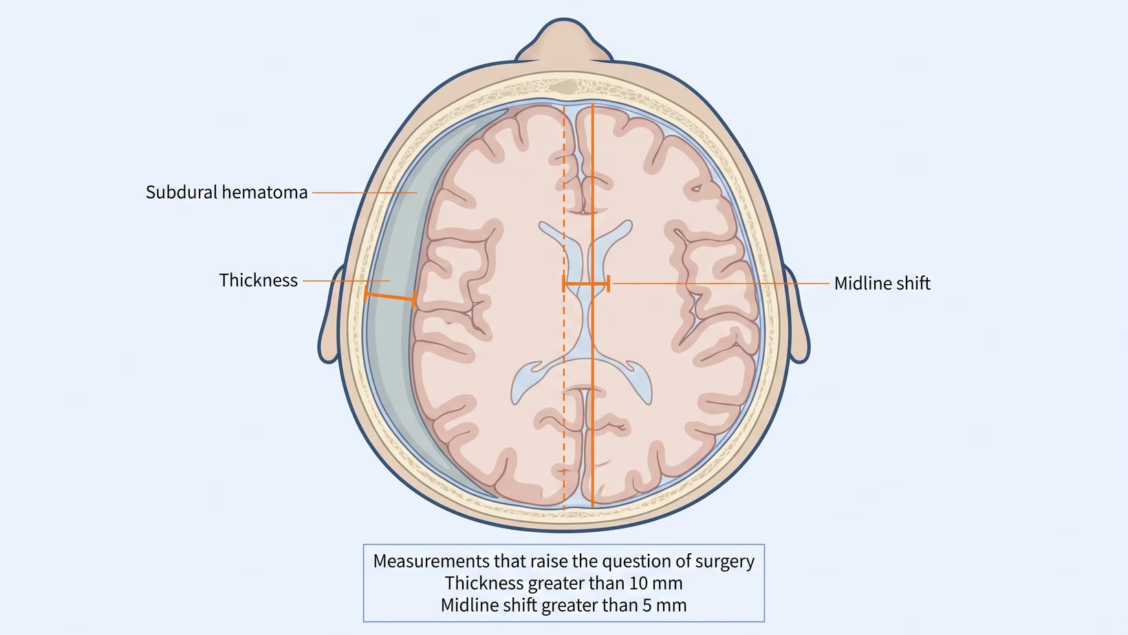
A small subdural hematoma is judged by two numbers from the CT scan. One is the thickness of the collection at its widest. The other is how far the midline, the center line between the two halves of the brain, has been pushed across. Below 10 mm of thickness and 5 mm of shift, with mild symptoms, watching becomes reasonable.
An older brain has lost volume, so a scan can look alarming while the person walks in talking normally. So the grading looks at the person as well as the scan.
Thickness and Midline Shift
For a chronic collection, surgery is the recommended treatment when thickness is greater than 10 mm, midline shift is greater than 5 mm, and the neurological symptoms are pronounced. Deciding on the measurement alone, whatever the person’s conscious level, is the rule for an acute bleed, where it is correct. In a stable person with a chronic collection there is no agreed cut-off. The measurements mean surgery should be considered. The symptoms decide it.
Three more findings point toward surgery in someone already in a coma with a smaller acute collection. The first is a drop of two or more points between injury and arrival on the Glasgow Coma Scale, a bedside score of how the person opens their eyes, speaks and moves. The others are unequal or fixed dilated pupils, and intracranial pressure, meaning pressure inside the skull, above 20 mm Hg.
When Is Observation Offered Instead of an Operation?
Observation instead of an operation is offered when symptoms are mild or absent and an operation carries real risk for that person. Reported deaths around the time of the operation reach about 11 in 100 older patients (11 percent). Surgery has its own dangers, so it is not chosen automatically.
Who Is Watched, and Who Is Operated On
Watched: a small collection, few or no symptoms, and high surgical risk from other illnesses. The scale used runs from no symptoms, through headache or an unsteady walk, to drowsiness with one-sided weakness, then stupor, meaning the person can barely be woken, and coma. Waiting and scanning is described for the mildest end of that scale.
Operated on: anyone with pronounced neurological symptoms, and more so once the thickness or the shift is past its cut-off.
How Long Does a Subdural Hematoma Take to Go Away?
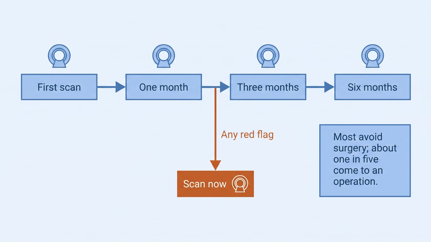
A subdural hematoma that is being watched usually takes months to go away. In the studies of people managed without an operation, the checks fall at one, three and six months from the first scan. Across studies, about 1 in 5 people managed this way still need surgery (18 to 20 percent).
What the Numbers Say, and What They Cannot Tell You
In a Dutch study of 83 people first treated without an operation, 61 needed nothing further within three months.
Which group you fall into is not predictable. A systematic review, a study that pools the results of many earlier studies, found no clinical or radiological sign showing which collection resolves on its own. Smaller collections do better as a group, but that cannot promise anything for one person. No study in the Philippines has measured this, so any percentage quoted to you comes from abroad.
Are There Medicines That Shrink a Subdural Hematoma?
Atorvastatin, a cholesterol medicine, is the medicine with the strongest trial evidence for shrinking a subdural hematoma. Dexamethasone, a steroid, was tested and did harm. Tranexamic acid is still being studied. None of them replaces the watching and the repeat scans.
Atorvastatin
Atorvastatin belongs to the statin group of medicines, and it was tested at a low dose. In a Chinese randomized trial of 196 patients treated without surgery, eight weeks of it reduced the collection by about 12.5 mL more than a dummy tablet (placebo). Surgery was needed by about 11 in 100 on atorvastatin (11.2 percent) against about 24 in 100 on placebo (23.5 percent), with no significant side effects reported.
Guidance supports it as an addition to observation, not a replacement for the scans. It is started only by the doctor treating the hematoma, who checks it against the person’s other medicines. Do not start it, or change the dose of a statin already taken, on your own.
Dexamethasone and Other Steroids
Dexamethasone was tested properly and the result went against it. Among 748 adults in a United Kingdom trial, a favorable outcome at six months was reached by about 84 in 100 on the steroid (83.9 percent) and about 90 in 100 on placebo (90.3 percent). Serious side effects happened in about 16 in 100 people on the steroid (16.0 percent), against about 6 in 100 on placebo (6.4 percent).
Almost all of those patients had surgery in the same admission, so the trial does not settle what steroids do in a collection that is only watched. It does show that this tablet is not harmless to add while waiting.
Tranexamic Acid
Tranexamic acid is a medicine that helps blood clot, and it has been studied mostly after surgery. Pooling six studies and 1,403 patients, it cut how often the collection came back after an operation by more than half. Guidance notes it can shrink a collection in selected cases, but it carries a risk of clotting elsewhere. It is not a tablet to buy and take on your own for this; only the treating doctor decides whether it fits, and whether it can spare someone an operation is still being tested.
Embolization of the Middle Meningeal Artery
Embolization means blocking a blood vessel from the inside. It is neither a tablet nor an open operation. A thin tube (catheter) is passed to the artery feeding the membrane around the collection and blocks its supply, so the membrane stops refilling it.
In a trial of 310 patients, adding embolization to standard treatment dropped treatment failure at 180 days to 16 in 100 (16 percent) from 36 in 100 (36 percent). Most of those patients were also having surgery; among the 121 who were not, the figures were about 19 in 100 (19 percent) against about 56 in 100 (56 percent).
Disabling stroke or death at 30 days happened in about 3 in 100 in both groups. Guidelines now recommend it alongside standard treatment, surgical or not, in selected non-emergency cases. Few centers here offer it.
What Should Be Stopped, and What Should Not
Blood thinners (anticoagulants and antiplatelet medicines) are part of why these collections form and enlarge. Stopping one on your own can cause a stroke or a clot, so that decision belongs to the prescribing doctor and the neurosurgeon together.
What Is the Repeat CT Scan Schedule, and What Does Each One Cost?
In the trials, a watched subdural hematoma is scanned again at one, three and six months from the first visit. A plain CT costs a different amount in each hospital, so ask radiology and the billing office before each scheduled scan. When a warning sign appears, go first and ask about cost after. The scan is how the watching is done, so it is not the place to save money.
How Often the Scan Is Repeated
For someone stable, with no symptoms or mild ones, guidance says less frequent imaging can be enough, with extra scans as needed if symptoms appear or worsen. A new symptom brings the scan forward and does not wait for the calendar.
What to Ask About the Cost
Prices for a plain CT of the head (cranial CT) change often, so ask rather than assume.
- Radiology, for the cash price of a plain cranial CT without contrast, and whether a follow-up scan is priced differently.
- The hospital’s PhilHealth or billing office, for what is covered in your case, and whether that changes if you are admitted. Their own case-rate search is at crs.philhealth.gov.ph.
- Radiology again, for your scan on a disc or as a file, so the next hospital compares images instead of repeating one.
When Does Observation Turn Into Surgery?
Observation turns into surgery when a repeat scan crosses a measurement the neurosurgeon uses, or when the person’s symptoms get worse. Either one is reason to reconsider surgery, and neither waits for the other. The measurements are a thickness above 10 mm and a midline shift above 5 mm.
The Thresholds a Neurosurgeon Uses
- Thickness greater than 10 mm on the repeat scan
- Midline shift greater than 5 mm
- Pronounced neurological symptoms, whatever the scan shows
Surgery is not a guaranteed end to the problem either. How often the collection comes back after it is drained (evacuation) varies widely between studies. A second operation is needed at 10 to 20 in 100 (10 to 20 percent), and the collection usually comes back within one to three months.
For the Referring Physician
Send the images rather than the report alone, because thickness and shift are measured from the images. Include the history of blood thinners (antithrombotics), the date of any fall, and the person’s baseline function before this illness.
Expect a decision framed as thresholds and a date rather than a yes or no. Confusion without a clear headache is the presentation most easily blamed on age or dementia, and it can appear weeks after a bump to the head.
Bring and ask
What to Bring and What to Ask
Bring:
- The CT images on a disc or file, not only the printed report
- The date and circumstances of any fall, even a minor one
- Every medicine and supplement, in their packaging
- Someone who sees the patient daily
Ask:
- How thick is it, and how far has the midline shifted
- What number would change the plan
- When is the next scan, and who do I call if something changes first
- What exactly should send us to the emergency room now
Frequently Asked Questions
Can I go back to work while we are watching it?
Going back to work while a subdural hematoma is watched depends on your symptoms and your job, so ask your own neurosurgeon. Work at heights, driving, and anything with a real chance of a bump to the head should wait.
Can a fresh bump on the head make it bleed again?
A new bump to the head means going to the nearest emergency room now to be scanned, even if the person seems well, as in the warning-sign list above.
My father is confused. Is that the bleed or is it dementia?
Confusion in an older person can come from a subdural hematoma or from dementia, and a new confusion is sometimes mistaken for dementia. Take it seriously. Confusion that is new or getting worse over hours or days means the nearest emergency room now. Confusion that has been the same for months still needs a doctor this week, who can check whether a scan is needed. Between 5 and 7 in 10 older patients with this condition arrive confused or less alert than usual (altered mental status).
Does this only happen to the elderly?
No, a subdural hematoma does not only happen to older people. It is most common over 65, and it is becoming more common as people live longer and take more blood thinners. It also occurs under 50. There the usual causes are bleeding disorders (coagulation disorders), arachnoid cysts (fluid-filled pouches on the lining of the brain), shunts (tubes that drain fluid from the brain) and injury. Headache and vomiting appear more often than one-sided weakness.
What to do now
What to Do Now
If a subdural hematoma is being watched, do these three things this week.
- Get the date of the next scan in writing, with a number to call if something changes first.
- Print the red-flag list above where whoever lives with the patient will see it.
- Book a review with a neurosurgeon, ideally the one who read the scan, within the week. Bring the CT images, every medicine in its packaging, and someone who sees the patient daily. You can book a consultation yourself, or your doctor can refer you.
Kung wala pa pong nakatakdang balikan, iyon po ang unang tatawagan ninyo bukas. Hindi po sapat ang basta paghihintay. Ang paghihintay na may petsa at may scan ang gamutan.
See sources
- Siddiq F, Shakir M, Nguyen TN, et al. Consensus Statement on Middle Meningeal Artery Embolization in Chronic Subdural Hematoma Treatment: A Guideline from the Society of Vascular and Interventional Neurology Guidelines and Practice Standards Committee. Stroke: Vascular and Interventional Neurology, 2025. PMID 41608698. DOI 10.1161/SVIN.125.001814
- Hutchinson PJ, Edlmann E, Bulters D, et al. Trial of Dexamethasone for Chronic Subdural Hematoma (Dex-CSDH). New England Journal of Medicine, 2020. PMID 33326713. DOI 10.1056/NEJMoa2020473
- Jiang R, Zhao S, Wang R, et al. Safety and Efficacy of Atorvastatin for Chronic Subdural Hematoma in Chinese Patients: A Randomized Clinical Trial (ATOCH). JAMA Neurology, 2018. PMID 30073290. DOI 10.1001/jamaneurol.2018.2030
- Fiorella D, Monteith SJ, Hanel R, et al. Embolization of the Middle Meningeal Artery for Chronic Subdural Hematoma (STEM). New England Journal of Medicine, 2024. PMID 39565980. DOI 10.1056/NEJMoa2409845
- Fakhry R, Dirven CMF, Moudrous W, et al. Additional treatment after primary conservative treatment in patients with chronic subdural hematoma: a retrospective study. Brain and Behavior, 2024. PMID 38956812. DOI 10.1002/brb3.3590
- Soleman J, Nocera F, Mariani L. The conservative and pharmacological management of chronic subdural haematoma. Swiss Medical Weekly, 2017. PMID 28102879. DOI 10.57187/smw.2017.14398
- Albalkhi I, Alaswad M, Saleh T, et al. Adjuvant Tranexamic Acid for Reducing Postoperative Recurrence of Chronic Subdural Hematoma in the Elderly: A Systematic Review and Meta-Analysis. World Neurosurgery, 2023. PMID 38101544. DOI 10.1016/j.wneu.2023.12.054
- Immenga S, Lodewijkx R, Roos YBWEM, et al. Tranexamic acid to prevent operation in chronic subdural haematoma (TORCH): study protocol for a randomised placebo-controlled clinical trial. Trials, 2022. PMID 35042560. DOI 10.1186/s13063-021-05907-0
- Pierre L, Julien C, Margetis K, Kondamudi NP. Subdural Hematoma. StatPearls Publishing, 2026. NBK532970
- Gillespie CS, Veremu M, Cook WH, et al. Middle meningeal artery embolization for chronic subdural hematoma: meta-analysis of three randomized controlled trials and review of ongoing trials. Acta Neurochirurgica, 2025. PMID 40493076. DOI 10.1007/s00701-025-06587-4