
Not every subdural hematoma needs surgery. A subdural hematoma is blood collecting between the brain and the skull. A fresh bleed after a head injury is an emergency, and when an operation is needed it is done as soon as possible. An old bleed is drained when it is large or causes symptoms. Small bleeds without symptoms are sometimes watched.
Does a subdural hematoma always need surgery?
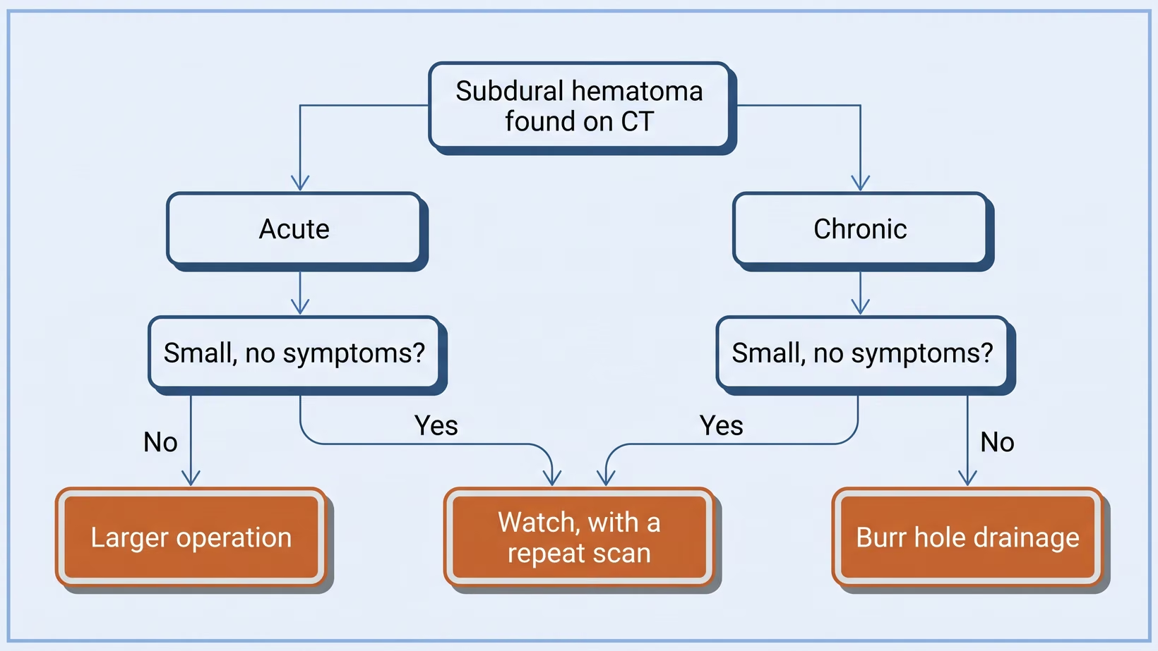
A subdural hematoma does not always need surgery. When the bleed is small and causes no symptoms, it is sometimes watched with a repeat scan. When it is large, or the person is confused, weak or getting drowsy, it is usually drained. Most patients are somewhere in between, and the decision is made with your neurosurgeon.
Acute or chronic? The one word that decides your treatment
An acute subdural hematoma is a fresh bleed, found in the hours after a definite head injury. It is an emergency, and the surgical guidelines say that when an operation is needed it should be done as soon as possible. A chronic subdural hematoma is an old bleed, found weeks after a bump. There is time to plan its treatment.
An acute clot is solid. In a chronic bleed, the bump may have seemed too small to matter, and by the time it is found the blood has turned to dark fluid.
What the doctors are measuring on your CT scan
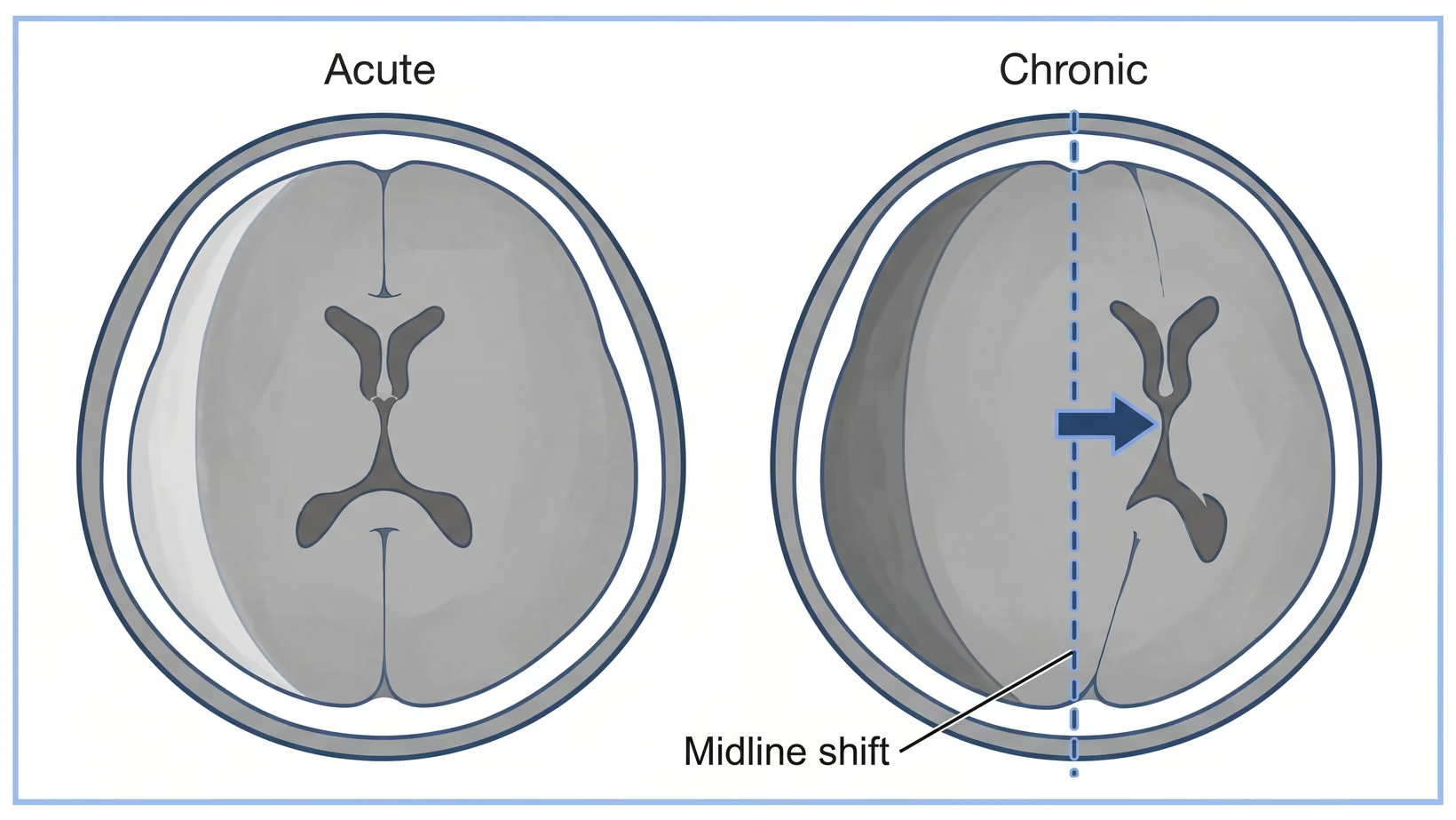
Doctors measure two things on the CT scan. One is the thickness of the clot at its widest point. The other is the midline shift. This is how far the middle of the brain has been pushed away from the middle of the skull by the pressure of the blood.
For an acute clot caused by an injury, the international surgical guidelines say it should be removed if it is thicker than 10 millimeters or the midline shift is more than five millimeters. This applies whatever the patient’s level of consciousness. It is a level above which surgery is needed. It does not mean a smaller clot is safe, and it is not a rule to apply to your own scan report at home.
For a chronic subdural hematoma, surgery is the main treatment for someone who has symptoms, or whose clot is pressing hard on the brain. So how the person is matters as much as the millimeters: whether they are confused, unsteady, weak on one side, or not themselves.
Seek care now
Warning signs that mean going to the emergency room now
Go to an emergency department straight away, and do not wait for a clinic appointment, if you or the person you are caring for has any of these:
- A headache that keeps getting worse instead of settling
- Vomiting more than once
- New weakness, heaviness or numbness in an arm or a leg, usually on one side
- Speech that has become slurred, or words that come out wrong
- New unsteadiness, or veering and falling toward one side
- A fit or seizure
- Growing drowsiness, or being hard to wake
- New confusion in an older person, especially if it came on over days or weeks
Symptoms that begin long after the injury are easy to blame on age or tiredness. The symptoms that start weeks after a bump on the head are the classic first signs of a slow bleed. This list is written for adults. For a baby or young child, go to the emergency room straight away if you are worried.
What does subdural hematoma surgery actually involve?
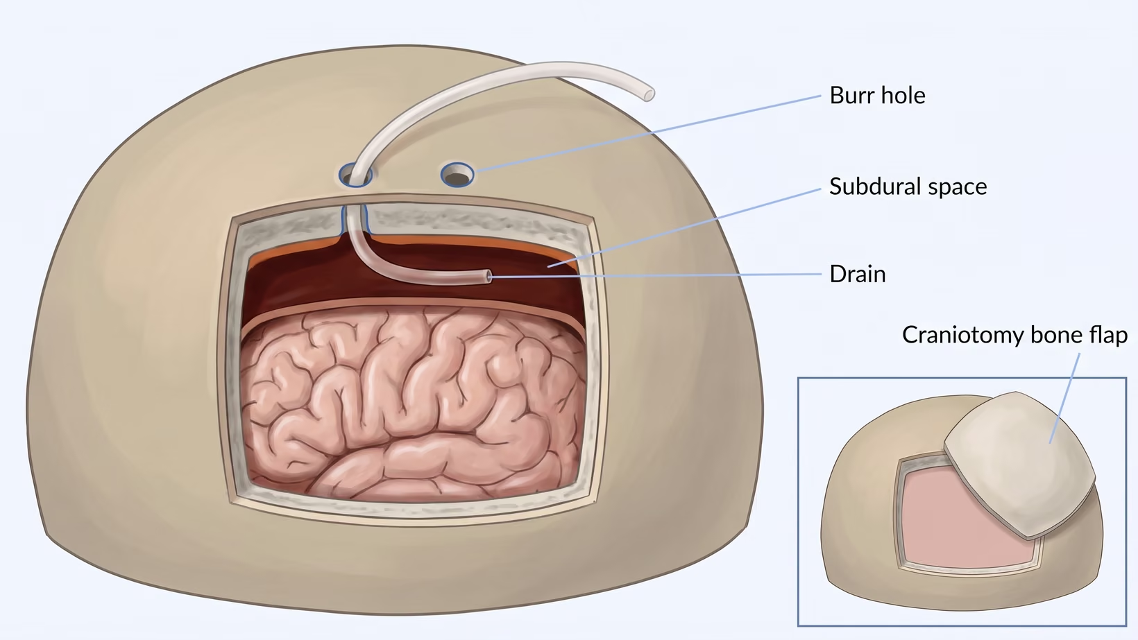
For a chronic subdural hematoma, the standard operation is burr hole drainage. The surgeon makes one or two small openings in the skull, called burr holes, and washes the dark fluid out through them. A soft drain tube is left in the space. A large acute clot needs a craniotomy, where a section of skull is removed.
Burr hole drainage does not always need a general anesthetic. It can be done with local anesthetic and sedation: the scalp is numbed, and the patient is kept drowsy and comfortable but not fully asleep. In a comparison between two Malaysian hospitals, most patients recovered well with either anesthetic. That study does not prove one method is better. Your surgeon and anesthesiologist choose, based on your age, your heart and lungs, and how still you can lie.
Solid clotted blood cannot be washed out through a small hole. In a craniotomy, the clot is removed and the bone is usually put back at the end. Sometimes the bone is left off on purpose to give the swollen brain room. This is called a decompressive craniectomy, and the bone is replaced at a later operation.
A trial of 450 patients with severe traumatic clots, who were assigned to either operation by chance, found that disability and quality of life a year later were similar with either operation. More patients needed further surgery after craniotomy, and more had wound problems after craniectomy. About three in ten had died by a year, which shows how serious a large traumatic bleed is.
Why the surgeon leaves a drain in after the operation
The drain is there to stop the bleed coming back. After the fluid is washed out, the space it filled does not close straight away, and it can fill again. A soft tube left in that space lets it keep emptying while the brain expands back into place. It lowers the chance of a second operation.
A randomized trial, meaning patients were assigned to each group by chance, studied 215 adults having burr hole surgery for a chronic subdural hematoma. The bleed came back in about 9 in every 100 patients who had a drain, and about 24 in every 100 without one.
Is there a newer procedure that stops it coming back?
There is a newer procedure called middle meningeal artery embolization, and the evidence for it is mixed. A thin tube (catheter) is passed from an artery in the wrist or groin to block the small artery that feeds the membranes around a chronic subdural hematoma. In trials it has mostly been done alongside surgery, not in place of it.
Two large trials reported in 2024 and did not agree. In the first, of 400 patients, the bleed came back and needed repeat surgery in about 4 in every 100 who had the embolization, and about 11 in every 100 who did not. In the second, of 722 patients in China, the difference at 90 days was too small to be sure it was real.
In the first trial the procedure caused a serious complication in about 2 in every 100 patients, including two disabling strokes. It also needs a specialist X-ray team (interventional neuroradiology).
Subdural hematoma medication: what medicines can and cannot do
No medicine has been shown to replace the operation for a large subdural hematoma that is causing symptoms. Two medicines have been tested to see whether a bleed can be made to shrink, or be less likely to come back. One is a steroid called dexamethasone. The other is a statin. Neither has replaced surgery.
In a trial of 748 adults with a chronic subdural hematoma causing symptoms, dexamethasone left fewer patients with a good outcome at six months than a placebo, a dummy treatment. About 84 in every 100 patients on the steroid had a good outcome, against about 90 in every 100 on placebo.
The steroid also caused more side effects. It did reduce repeat operations, from about 7 in every 100 on placebo to about 2 in every 100 on the steroid. Most of these patients also had surgery, so the steroid was given alongside the operation.
A statin is one of the common medicines that lower cholesterol. In an early trial of 196 patients in China, all being managed without surgery, eight weeks of a statin shrank chronic subdural hematomas more than placebo. Fewer of those patients needed an operation during the trial. It is still research, not yet a treatment to ask for.
Do not start or stop a medicine on your own. If you are already on a steroid for this, your team chose it with your scan in front of them. If this worries you, ask them before your next dose and do not stop it before they answer.
What happens to my blood thinner or aspirin?
There is no single agreed rule for a blood thinner or aspirin after a subdural hematoma, so your team decides it for you. They weigh two real risks. If the medicine continues, the bleed can grow or come back. If it stops, there is a risk of a stroke, a heart attack or a blood clot.
A review found no randomized trials and no guideline that settles low-dose aspirin in chronic subdural hematoma. The balance depends on why you take the medicine in the first place.
Before you leave the hospital, ask when to stop and when to restart the blood thinner, and who to call in between. Never stop a blood thinner on your own, and never restart one on your own either.
What if the bleed is small and I feel well?
If the bleed is small and you feel well, an operation may not be the first step. A small subdural hematoma that causes no symptoms can be watched instead of operated on, with a repeat scan to see which way it is going. The decision is then made on what that scan shows.
Being watched is a treatment plan. It only works if the repeat scan is done. If any of the warning signs above appear while you are waiting for that scan, go to the emergency room now.
How long does recovery take, and what should I expect?
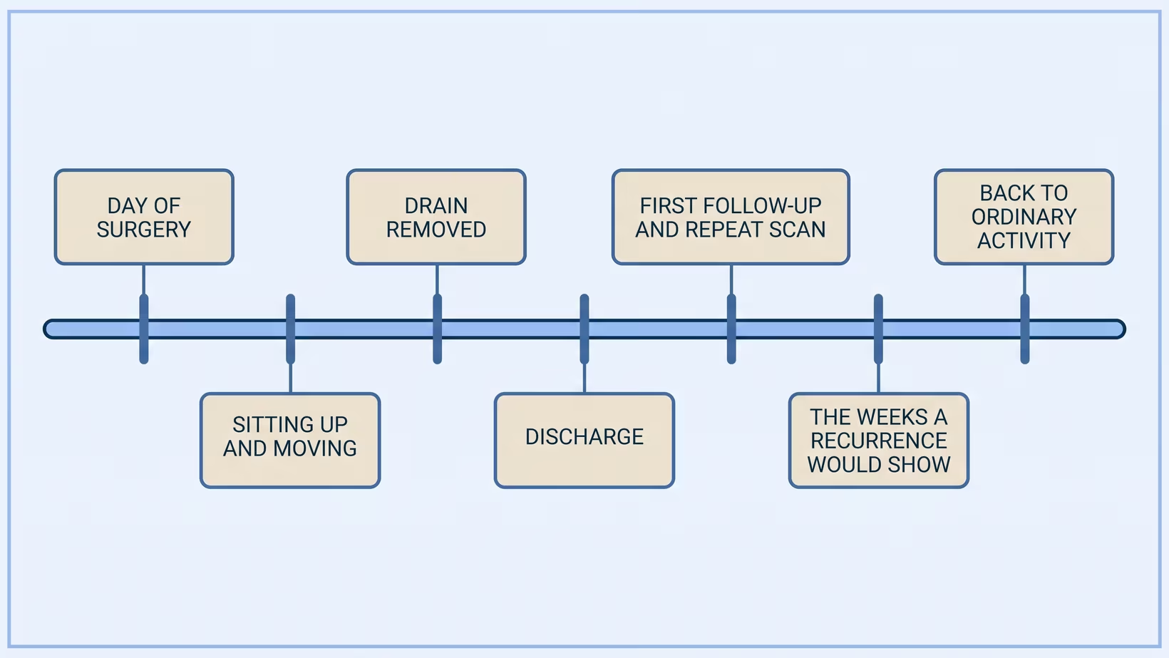
Recovery from burr hole drainage differs from person to person. The drain comes out after a short time, and the wound is small. Older patients can become clearer once the pressure is off the brain. Your team arranges a follow-up scan, and its timing depends on the hospital and on how you are doing.
Do not expect to be kept flat in bed. A trial in Portugal compared patients whose head of bed was raised within 12 hours after burr hole surgery with patients kept flat for two days. A year later, more of the early group were functioning well, and their bleed came back no more often. This does not mean getting up alone or before you are asked. When the ward team wants the patient sitting up and moving, follow that instruction.
Recovery after a craniotomy for a large acute clot depends far more on the brain injury than on the wound.
Can it come back after surgery?
Yes, a chronic subdural hematoma can come back after surgery in a minority of patients. Across published studies, it comes back in between 5 and 30 in every 100 patients. The range is wide because studies counted in different ways, so it is not a prediction for any one person.
A drain left in after burr hole surgery lowers that chance. Blocking the artery that feeds the membranes may lower it further, though the two trials of it disagree.
Watch for the same symptoms that brought you in. A headache getting worse again, new confusion, new weakness or new drowsiness in the weeks after surgery means going to the emergency room now. Do not wait for the follow-up appointment.
What does treatment cost, and what does PhilHealth cover?
The cost of subdural hematoma surgery depends on the hospital, on whether it was a burr hole or a craniotomy, and on how many days are spent in the ward or in intensive care. Repeat scans and complications add to it, and PhilHealth case rates change. So no figure is given here.
Ask the billing section for the PhilHealth case rate for this admission and this operation, what it is expected to leave unpaid, and whether the neurosurgeon, anesthesiologist and radiologist bill separately. Ask to speak to the medical social worker about the assistance that hospital can process. Do this early, and send someone who is not the patient.
What to do now
What to do next: who to see and what to bring
If a scan has already shown a subdural hematoma, the person to see is a neurosurgeon. The referral is arranged by whoever ordered the scan: your family doctor, your internist, or the emergency physician who saw you. Ask for it now rather than waiting to see whether the symptoms settle. You can also book a consultation yourself. If any of the warning signs above appear, go to the emergency room instead of waiting for the referral.
If there has been no scan yet, and the concern is an older person who has had a fall and hit the head, or someone who hit their head while taking a blood thinner or aspirin, see a doctor about it soon. Not everyone needs an urgent consult, but if the person feels unwell, or any of the warning signs above appear (a worsening headache, vomiting more than once, new weakness, confusion or drowsiness, or a seizure), go to the emergency room straight away to be checked.
If you would like me to see you, I see adults at my clinic at the Lung Center of the Philippines, Room 1101, Quezon Avenue, Quezon City, on Tuesday and Thursday from 2 PM. If the trip to Quezon City is not practical, you can book an online consult on the consultation page.
Bring and ask
What to bring, and what to ask
Bring to the consultation:
- The actual CT films or the disc, and the radiologist’s written report, not a photograph of the screen
- A written list of every medicine, including aspirin, any blood thinner, and anything bought over the counter
- The date of the fall or the bump, even if it seemed trivial at the time
- Someone who was there, because the patient often does not remember it
Ask the neurosurgeon:
- Is this bleed acute or chronic, and how thick is it
- Is the brain being pushed across, and by how much
- If we watch this instead of operating, what exactly are we watching for
- If we operate, would it be a burr hole or a craniotomy, and what anesthetic
- What would tell us it has come back, and who do I call
Frequently asked questions
Is subdural hematoma surgery done under general anesthesia?
Subdural hematoma surgery is not always done under general anesthesia. Burr hole drainage for a chronic subdural hematoma can be done with local anesthetic and sedation, which keeps the patient drowsy but not fully asleep. A craniotomy for a large acute clot is done under general anesthesia. Your surgeon and anesthesiologist decide which is right for you.
Can medicine dissolve a subdural hematoma instead?
No medicine has been shown to take the place of surgery for a large subdural hematoma that is causing symptoms. Dexamethasone, a steroid, reduced repeat operations but left fewer patients with a good outcome. A statin shrank bleeds in one early trial of patients managed without surgery, and that work is still at an early stage.
When can I restart my blood thinner?
There is no single agreed rule for restarting a blood thinner after a subdural hematoma. For aspirin, a review found no trial or guideline that settles it. Your surgeon decides with the doctor who prescribed it, weighing your risk of another bleed against your risk of a clot. Leave the hospital knowing the plan and the date.
How soon can someone get out of bed after the operation?
After burr hole surgery for a subdural hematoma, patients usually get out of bed sooner than families expect, on the ward team’s timing. In a trial, patients whose head of bed was raised within 12 hours were doing better a year later than those kept flat. Their bleed came back no more often.
Will it come back?
A chronic subdural hematoma comes back after surgery in a minority of patients: between about 5 and 30 in every 100 across published studies. A drain left in after burr hole surgery lowered it from about 24 in 100 to about 9 in 100 in one trial. If the symptoms that first brought you in come back, go to the emergency room straight away.
See sources
- Bullock MR, Chesnut R, Ghajar J, et al. Surgical management of acute subdural hematomas. Neurosurgery, 2006. PMID 16710968.
- Santarius T, Kirkpatrick PJ, Ganesan D, et al. Use of drains versus no drains after burr-hole evacuation of chronic subdural haematoma: a randomised controlled trial. The Lancet, 2009. PMID 19782872 / 10.1016/S0140-6736(09)61115-6.
- Wong HM, et al. Chronic Subdural Hematoma Drainage Under Local Anesthesia with Sedation versus General Anesthesia and Its Outcome. World Neurosurgery, 2021. PMID 34648987 / 10.1016/j.wneu.2021.10.074.
- Al-Salihi MM, et al. Comparison of Burr-Hole Craniostomy versus Twist-Drill Craniostomy Operations for Patients with Chronic Subdural Hematoma: A Systematic Review and Network Meta-Analysis. World Neurosurgery, 2023. PMID 37178912 / 10.1016/j.wneu.2023.05.022.
- Hutchinson PJ, Edlmann E, Bulters D, et al. Trial of Dexamethasone for Chronic Subdural Hematoma. New England Journal of Medicine, 2020. PMID 33326713 / 10.1056/NEJMoa2020473.
- Jiang R, Zhao S, Wang R, et al. Safety and Efficacy of Atorvastatin for Chronic Subdural Hematoma in Chinese Patients: A Randomized Clinical Trial. JAMA Neurology, 2018. PMID 30073290 / 10.1001/jamaneurol.2018.2030.
- Davies JM, Knopman J, Mokin M, et al. Adjunctive Middle Meningeal Artery Embolization for Subdural Hematoma. New England Journal of Medicine, 2024. PMID 39565988 / 10.1056/NEJMoa2313472.
- Liu J, Ni W, Zuo Q, et al. Middle Meningeal Artery Embolization for Nonacute Subdural Hematoma. New England Journal of Medicine, 2024. PMID 39565989 / 10.1056/NEJMoa2401201.
- Hutchinson PJ, Adams H, Mohan M, et al. Decompressive Craniectomy versus Craniotomy for Acute Subdural Hematoma. New England Journal of Medicine, 2023. PMID 37092792 / 10.1056/NEJMoa2214172.
- Pinto V, et al. GET-UP Trial 1-year results: long-term impact of an early mobilization protocol on functional performance after surgery for chronic subdural hematoma. Journal of Neurosurgery, 2023. PMID 37948693 / 10.3171/2023.8.JNS231509.
- Mongardi L, et al. Low-Dose Acetylsalicylic Acid in Chronic Subdural Hematomas: A Neurosurgeon’s Sword of Damocles. Frontiers in Neurology, 2020. PMID 33133003 / 10.3389/fneur.2020.550084.
- Stubbs DJ, et al. Incidence of chronic subdural haematoma: a single-centre exploration of the effects of an ageing population with a review of the literature. Acta Neurochirurgica, 2021. PMID 34181085 / 10.1007/s00701-021-04879-z.
- Kolias AG, Chari A, Santarius T, Hutchinson PJ. Chronic subdural haematoma: modern management and emerging therapies. Nature Reviews Neurology, 2014. PMID 25224156 / 10.1038/nrneurol.2014.163.