

A slow bleed under the skull can grow for weeks after a fall. Go to the emergency room now if he was knocked out, cannot remember the fall, is confused or drowsy, vomits repeatedly, has a severe headache, one-sided weakness, slurred speech, a drooping face or a seizure, or takes a blood thinner. Otherwise see a doctor today.
Lolo Fell in the Bathroom. What Happens Now?
First, look at him right now. Is he awake, talking the way he always talks, moving both sides of his body, and does he remember falling? If the answer to any of these is no, he goes to the nearest emergency room now. If yes, read the list below before deciding anything. If anything on it is true, or he takes a blood thinner, he goes to the emergency room now. If nothing on it is true, have him seen by a doctor today, because at his age a scan is often advised even when he looks well.
Huwag po kayong mag-panic, pero huwag din ninyong kalimutan. Falls at this age are ordinary rather than freakish. In the Longitudinal Study of Ageing and Health in the Philippines, a nationally representative survey of 4,606 older Filipinos, 17.7 percent had fallen in the year before they were asked.
In a Japanese registry of 1,445 people with a chronic subdural hematoma (a slow collection of blood under the brain’s outer covering, between the brain and the skull), a ground-level fall was the most common cause among older patients. A traffic accident was the most common cause among younger ones. The fall that looks least serious is the one that produces this. That is what a chronic subdural hematoma is and why elders get it.
Seek care now
Which Signs Mean the Emergency Room Now?
Any one of these means going now to the nearest emergency department with a CT scanner (the machine that takes pictures of the brain), not waiting for tonight and not a clinic on Monday.
- He was knocked out, even for a few seconds
- He cannot remember the fall itself
- He is confused or drowsy in a way he was not this morning
- Repeated vomiting
- A severe headache, or one that keeps getting worse
- Weakness on one side, slurred speech, or a drooping face
- A seizure
- Clear fluid or blood from the nose or the ear
- One pupil bigger than the other
- He takes a blood thinner and he struck his head at all
Bring the pill box, not a list from memory. If he gets worse on the way, that is the picture of the acute subdural hematoma, the one where hours decide.
He Hit His Head but Seems Fine. Is That Enough?
Seeming fine is real reassurance about tonight and says very little about next month. The two run on different clocks, and that is the whole reason this page exists.
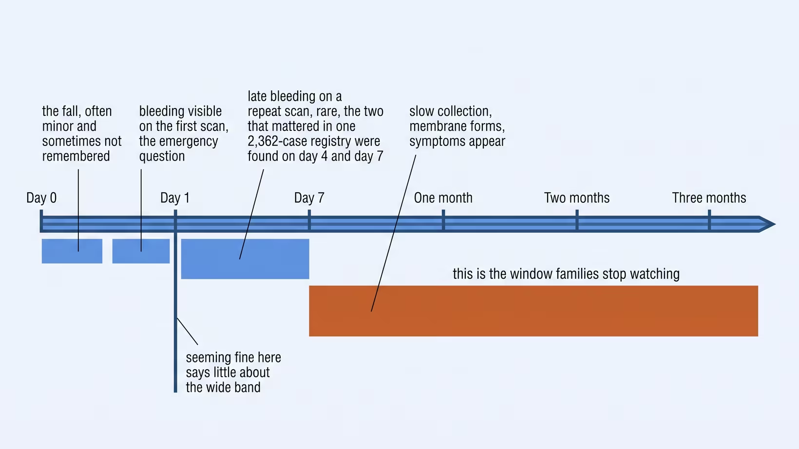
The slow bleed does not cause early symptoms. In the reviewed literature, the great majority of people who are found to have a chronic subdural hematoma arrive alert and near-normal, weeks after the bump that started it. Across the published series, somewhere between a quarter and half of the people found to have one recall no head injury at all, or only a bump too small to have thought about.
So what about the first day? A Swedish registry study of 2,362 head injuries in people on blood thinners, median age 84, found bleeding that appeared only on a later scan in five patients, and only two of those, about 1 in 1,000 (0.08 percent), mattered clinically. Most of the risk of a late bleed does not sit in that first night, but the first night still matters. Even so, if the hospital asks him to stay, stay. The guideline those researchers were arguing with still asks for 24 hours of watching after a head injury on a blood thinner, and they were calling for it to be reconsidered rather than reporting that it had changed.
Read the rest of that study before relaxing. Those two late bleeds were both subdural, they were found on day four and day seven, and both patients died. The window to watch is weeks long, and the trigger to come back is a change in him rather than a date. These are symptoms that appear weeks after a bump on the head.
Why Does an Elder Get a Scan Even Without Symptoms?
Because age moves the odds far enough that the usual warning signs stop working as a filter. What reliably sorts a 40-year-old does not reliably sort an 85-year-old.
Among 4,308 patients aged 65 and over who reached emergency departments in Canada and the United States after a ground-level fall, median age 83, 139 of them, or 3.2 percent, had clinically important bleeding inside the skull. About one in 31 of everyone who walks in, including plenty who look entirely well.
Those researchers derived a Falls Decision Rule to spare some of them the radiation: no CT needed if there was no head injury in the fall, no amnesia for it, no new abnormality on neurological examination, and a score below five on the Clinical Frailty Scale, which rates how frail a person is. It caught 98.6 percent of the bleeds. Their own paper states the rule still needs testing in other hospitals before it guides decisions, which is exactly why the threshold for scanning an older person stays low in practice. The rule is a tool for emergency doctors, not one a family can use at home to decide against a scan.
Blood thinners are the one modifier a family can check themselves. In a Danish study of 10,010 people with a subdural hematoma matched against the general population, 47.3 percent were on an antithrombotic drug. Adjusted odds of a subdural hematoma ran at 1.24 with low-dose aspirin, 1.87 with clopidogrel and 1.73 with a direct oral anticoagulant (a blood thinner taken by mouth), rising to 3.69 with a vitamin K antagonist such as warfarin and higher still when that was combined with an antiplatelet (a blood thinner such as aspirin, which stops the platelets, the blood’s clotting cells, from sticking together).
This appears at the hospital door. In a prospective series of anticoagulated patients with a minor head injury, about 11 in 100 (11.2 percent) already had bleeding on the first scan. Of those on a vitamin K antagonist, about 16 in 100 (15.7 percent) had bleeding on the first scan, against about 5 in 100 (4.7 percent) of those on a direct oral anticoagulant. Say the words “blood thinner” at triage. It changes the queue.
Brain Shrinkage and the Bridging Veins
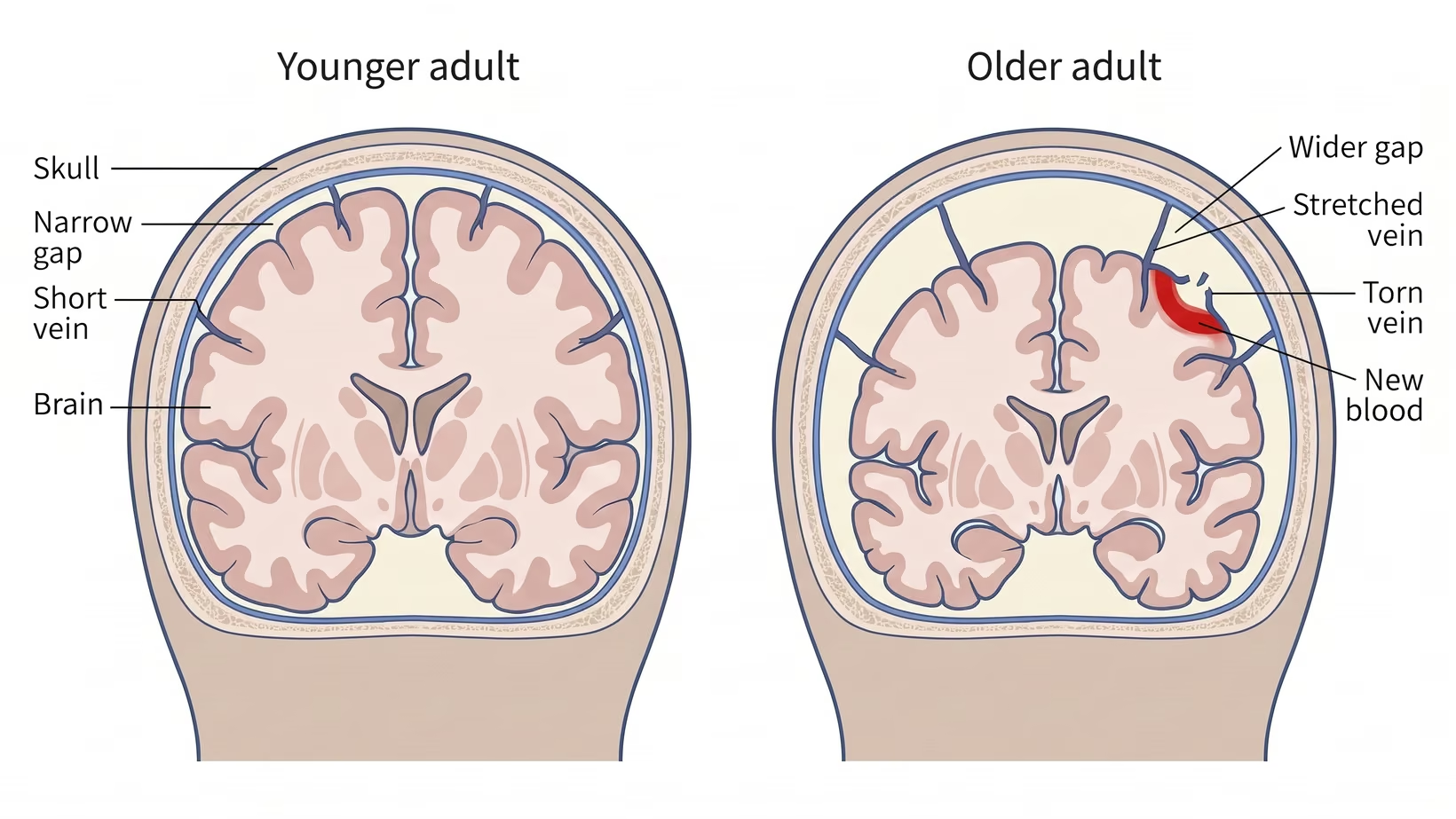
The brain loses volume with age while the skull stays the size it always was. The small veins crossing that widening gap are stretched, and a bump a younger head absorbs can tear one.
The mismatch has a name: craniocerebral disproportion. The bleeding is believed to come from those bridging veins where they cross the dura, the tough membrane that covers the brain, at the dural border cell layer, the thin plane between the brain’s coverings, where their walls can be as little as a single layer of cells. The same disproportion stretches the veins and leaves room for a collection to grow, with less brain pushing back against it.
That is why nothing happens for weeks. The blood is low-pressure venous blood seeping into a potential space, so the collection builds slowly and the symptoms arrive long after the injury. Reviewers describe three phases: a trauma that can pass unnoticed, weeks to months of quiet enlargement while a membrane forms around the clot, then the point where the skull runs out of room and the person finally changes.
The evidence has a limit. Those same reviewers call the literature on craniocerebral disproportion limited, so treat it as the best current explanation rather than a closed case.
What to Watch For in the Next Month
Most of the time you are not watching for a collapse. You are watching for a slow change in the person, and the people who live with him will see it before any test does. A sudden drop in alertness or a seizure still means the emergency room now.
The classic three are confusion, headache and drowsiness, with a seizure in a smaller group. In a Japanese national series of 63,358 patients, more than half of those aged 70 and over already had some disturbance of consciousness by the time they reached hospital.
At home it looks like this:
- New unsteadiness, or a change in how he walks
- Weakness in one arm or one leg, more common in the arm in one surgical series
- Falling more often than he used to
- Taking longer to answer, or losing the thread
- Sleeping through the day
- A headache that is new for him
- A personality the family describes as not quite him
Any one of these, new since the fall, means he is seen by a doctor today and a CT scan is asked for, even if the fall was weeks ago and the first scan was clear. Weakness on one side, a seizure, drowsiness or confusion that is getting worse by the hour, or drowsiness you cannot wake him from means the nearest emergency room now.
A chronic subdural hematoma is described in the literature as one of the recognized causes of reversible dementia, and in a North Wales series falls and confusion together were the reason for 90 percent of the scans that found one. A family that files the change under matanda na kasi files away the reversible thing. How doctors tell a chronic subdural hematoma from dementia is explained on its own page.
Bring and ask
What to Bring and What to Ask
Bring:
- The pill box itself, including anything bought over the counter
- A written list of his other conditions
- The date and story of the fall, written down, because he may not remember
- His usual self in concrete terms: who he recognizes, how far he walks, what he does by himself
- Any previous CT or MRI, on a disc
Ask:
- Does he need a CT today, and does the blood thinner change that answer
- If the scan is clear, what do we watch for and for how long
- What would bring us straight back
- Is his blood thinner continued, paused or changed, and who decides
- What does the scan cost here, and is any part of it covered
That last question carries no number here on purpose. Prices and coverage differ between hospitals and change over time, so ask the clerk and the doctor rather than a figure found online.
When the Family Should Not Wait
Stop waiting the moment you notice he is not himself and catch yourselves explaining it away. That sentence, not a symptom, is the signal. Have him seen by a doctor that same day and ask for a CT scan; if he is drowsy, weak on one side or has a seizure, go to the emergency room now.
This is a different failure from the red flags above. Nobody missed the sign. They saw it and decided it could wait until after the fiesta, after payday, or until the appointment already booked for next month.
What that costs has been measured. In an Italian series of fall-related subdural hematomas, patients who already had dementia or parkinsonism waited significantly longer between the trauma and admission and arrived more disabled, and unlike every other group in that study they did not measurably improve during the admission. In a separate surgical series, patients whose weakness had lasted longer before the operation regained strength less often.
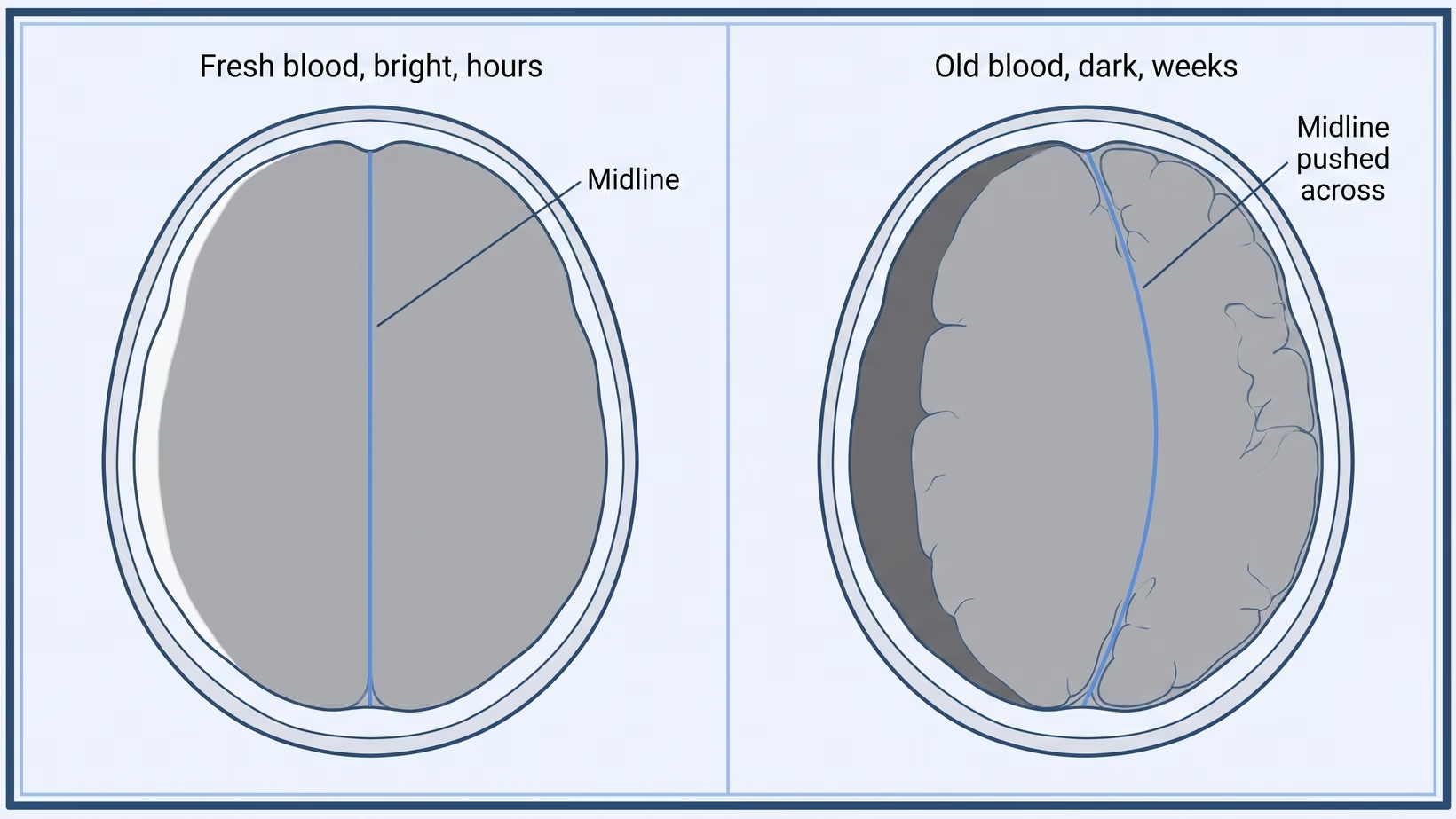
Now both halves of the outcome, because a family pushing for a scan deserves both. About 72 in 100 patients in the Japanese national series (71.6 percent) were independent in day-to-day life by the time they left hospital, and about two-thirds in that surgical series regained strength. Against that, Finnish national data put one-year case fatality after the operation at 15 percent, and a separate Finnish study found more deaths within one year in patients who already had a diagnosis of dementia. Surgery is offered when the collection is causing symptoms, or is thick, or is pushing the midline, the center line of the brain, across. Ito po ang karaniwang ginagawa, and whether it fits your father is a conversation with the surgeon who has seen his scan. There are also situations where a subdural hematoma can be watched rather than drained.
Frequently Asked Questions
Matanda Na Naman Po Kasi. Kailangan Pa Ba ng CT Scan?
Age is not a reason not to perform a scan. Among people 65 and over seen after a ground-level fall, about 3 in 100 (3.2 percent) had important bleeding inside the skull.
Does a Normal CT Scan Mean He Is in the Clear?
It means there is no bleed today, which is the question the scan was asked. A chronic subdural hematoma forms over weeks, so a clear scan on day one does not cover day 30.
He Takes Aspirin. Is That the Same as a Blood Thinner Here?
Count it, yes, and say so at triage. Do not stop it on your own; ask the doctor who prescribed it. Low-dose aspirin carried higher adjusted odds of subdural hematoma in the Danish study, lower than warfarin but not zero.
Can a Chronic Subdural Hematoma Go Away by Itself?
Small collections causing no symptoms are sometimes watched rather than drained. That call rests on the size, the pressure on the brain and how he is doing, and it belongs to the doctor reading the scan.
How Long Should We Keep Watching Him?
Watch him closely tonight against the red-flag list above, then keep ordinary attention for a month. The late bleeds that mattered in the Swedish registry turned up on day four and day seven.
Are There Philippine Numbers for This?
Not for this condition. National neurotrauma data is thinnest in the countries carrying the heaviest burden, so the figures here come from abroad and the one Philippine number is the falls figure above.
What to do now
What to Do Now
If anything on the red-flag list is true, go to the emergency room now. Otherwise have him seen by a doctor today and ask for a CT scan. If a collection is found, his doctor can refer him to a neurosurgeon, or you can book a consultation yourself. Bring the pill box and the date of the fall.
If you would like me to be the neurosurgeon who looks at his scan, my clinic is at the Lung Center of the Philippines, Room 1101, Quezon Avenue, Quezon City, on Tuesday and Thursday from 2 PM.
If he lives far away, or the trip is too hard for him, book an online consult on the consultation page.
Tonight, read the red-flag list out loud to whoever is staying with him, so two people know it instead of one.
Write the date of the fall on the calendar and leave it there for two months. The most useful thing you can hand a doctor in November is that he fell in September.
Photograph the pill box and put it where the family group chat can find it at midnight.
Fix the bathroom this week: a grab bar beside the toilet and inside the shower, a light that stays on all night, and the sliding mat taken out.
If he is on a blood thinner, ask his own doctor now, before anything happens, what the plan is the next time he falls.
Karamihan po ng pagkadapa ay walang masamang kasunod. Ang trabaho ninyo sa susunod na buwan ay ang mapansin, hindi ang mag-alala.
See sources
- Mgabhi PS, Chen TY, Cruz G, Vu NC, Saito Y. Falls among community-dwelling older adults in the Philippines and Viet Nam: results from nationally representative samples. Injury, 2024. PMID 38350305. DOI 10.1016/j.injury.2024.111336
- Karibe H, Kameyama M, Kawase M, Hirano T, Kawaguchi T, Tominaga T. Epidemiology of chronic subdural hematomas. No Shinkei Geka, 2011. PMID 22128269
- Adhiyaman V, Chattopadhyay I, Irshad F, Curran D, Abraham S. Increasing incidence of chronic subdural haematoma in the elderly. QJM, 2017. PMID 28069915. DOI 10.1093/qjmed/hcw231
- de Wit K, Mercuri M, Clayton N, et al. Derivation of the Falls Decision Rule to exclude intracranial bleeding without head CT in older adults who have fallen. CMAJ, 2023. PMID 38049159. DOI 10.1503/cmaj.230634
- Gaist D, García Rodríguez LA, Hellfritzsch M, et al. Association of antithrombotic drug use with subdural hematoma risk. JAMA, 2017. PMID 28245322. DOI 10.1001/jama.2017.0639
- Cipriano A, Pecori A, Bionda AE, et al. Intracranial hemorrhage in anticoagulated patients with mild traumatic brain injury: significant differences between direct oral anticoagulants and vitamin K antagonists. Internal and Emergency Medicine, 2018. PMID 29520701. DOI 10.1007/s11739-018-1806-1
- André L, Björkelund A, Ekelund U, Vedin T, Björk J, Forberg JL. The prevalence of clinically relevant delayed intracranial hemorrhage in head trauma patients treated with oral anticoagulants is very low. Scandinavian Journal of Trauma, Resuscitation and Emergency Medicine, 2024. PMID 38730480. DOI 10.1186/s13049-024-01214-0
- Nouri A, Gondar R, Schaller K, Meling T. Chronic subdural hematoma (cSDH): a review of the current state of the art. Brain and Spine, 2021. PMID 36247395. DOI 10.1016/j.bas.2021.100300
- Bin Zahid A, Balser D, Thomas R, Mahan MY, Hubbard ME, Samadani U. Increase in brain atrophy after subdural hematoma to rates greater than associated with dementia. Journal of Neurosurgery, 2018. PMID 29498578. DOI 10.3171/2017.8.JNS17477
- Toi H, Kinoshita K, Hirai S, et al. Present epidemiology of chronic subdural hematoma in Japan: analysis of 63,358 cases recorded in a national administrative database. Journal of Neurosurgery, 2017. PMID 28156246. DOI 10.3171/2016.9.JNS16623
- Nisson PL, Francis J, Michel M, et al. Focal motor weakness and recovery following chronic subdural hematoma evacuation. Journal of Neurosurgery, 2024. PMID 38875718. DOI 10.3171/2024.4.JNS24121
- Arca R, Ricchi V, Murgia D, et al. Parkinsonism and dementia are negative prognostic factors for the outcome of subdural hematoma. Neurological Sciences, 2016. PMID 27120071. DOI 10.1007/s10072-016-2588-8
- Sahyouni R, Goshtasbi K, Mahmoodi A, Tran DK, Chen JW. Chronic subdural hematoma: a perspective on subdural membranes and dementia. World Neurosurgery, 2017. PMID 28935547. DOI 10.1016/j.wneu.2017.09.063
- Tommiska P, Luostarinen T, Kaprio J, et al. Incidence of surgery for chronic subdural hematoma in Finland during 1997-2014: a nationwide study. Journal of Neurosurgery, 2021. PMID 34507291. DOI 10.3171/2021.3.JNS21281
- Tommiska P, Korja M, Siironen J, Kaprio J, Raj R. Mortality of older patients with dementia after surgery for chronic subdural hematoma: a nationwide study. Age and Ageing, 2021. PMID 33022060. DOI 10.1093/ageing/afaa193
- Thango N, Baticulon RE, Lippa L. The role of registries in neurotrauma research: translating data into health policy that enhances patient care. International Journal of Health Policy and Management, 2023. PMID 37579434. DOI 10.34172/ijhpm.2023.7521