
> A meningioma is a tumor that grows from the meninges, the layers that cover the brain, rather than from brain tissue itself. It is the most common primary brain tumor, and most are not cancer. Many are found by accident on a scan and if small, are monitored rather than removed.
What does meningioma mean?
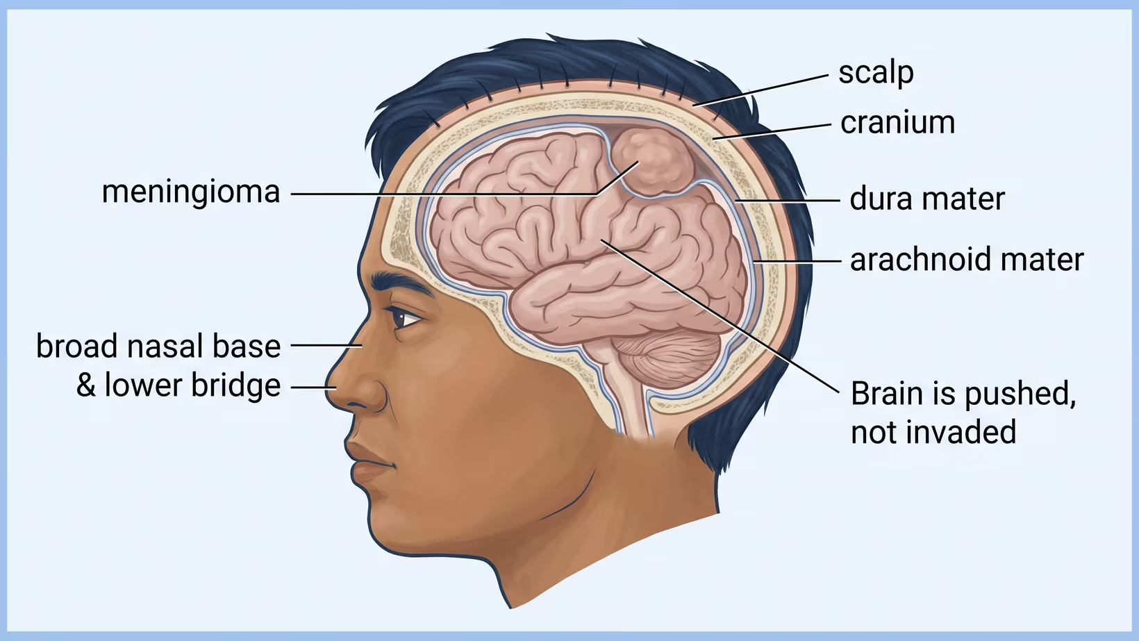
The word joins two parts. Meninges are the covering layers wrapped around the brain, and the ending oma means a growth. So a meningioma is a growth that starts in the covering, not in brain tissue.
Those coverings have names you will see in a report. The dura is the tough outer layer that lines the inside of the skull. The arachnoid is the thin, delicate layer beneath it. Under that is a space filled with fluid, and then the pia, a very fine layer lying on the brain itself. A meningioma grows from these layers and sits on the brain, pressing on it from outside.
That single fact explains most of what follows. A tumor that starts outside the brain can often be separated from it. It also means the trouble it causes usually comes from pressure and from what sits next to it. A minority grow into the brain surface, and when they do the pathologist grades them higher.
In most people, no cause is ever identified. A few things are known to raise the risk: radiation to the head in the past, long-term use of certain high-dose hormone medicines, and some inherited conditions that run in families. Tell your doctor about any of these.
Is a meningioma a benign brain tumor, or is it cancer?
Most meningiomas are not cancer. Benign describes how the cells look and behave under a microscope, and it is not the same as harmless, because a slow growth in a closed space can still cause problems.
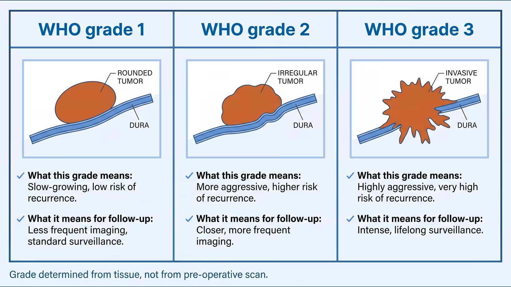
The grade is the number that matters, and it does not come from the scan. It comes from tissue examined by a pathologist under an international World Health Organization standard. Since 2021 that standard combines what is seen under the microscope with molecular testing, reported together as one integrated diagnosis. Molecular testing is not offered at every centre in the Philippines, so your report may or may not carry it.
Among meningiomas found by accident and then operated on, the great majority prove to be the slowest growing grade. A minority are more active and need closer follow-up. As a group, tumors that are not malignant carry a far better outlook than malignant brain tumors do.
No page can tell you which grade yours is. That is what the imaging plus the tissue answer.
How fast does a meningioma tumor grow?
Most meningiomas grow slowly, and some do not grow measurably at all. Growth is not judged from one scan; it is judged by comparing a new scan against an earlier one, which is why a repeat scan is often the next step rather than an operation.
About one in three meningiomas is discovered by accident, on a scan done for another reason. Among those watched rather than treated, only a small minority go on to develop symptoms. About a quarter eventually have some treatment, and on average that comes around two years after the tumor was found.
Two features mark out the ones that declare themselves: a larger tumor, and swelling in the brain around it. When a watched meningioma does change, it usually does so within the first five years after diagnosis.
This is why old scans are worth hunting down. A film from four years ago turns a guess into a measurement.
What symptoms does a meningioma cause?
Many meningiomas cause no symptoms at all and are found when a scan is done for something else. When symptoms do appear, they usually build up gradually over weeks or months rather than arriving in an instant.
What people notice most often:
- Headache that is new, or different from the headaches they are used to
- A seizure, which is one of the recognised ways a meningioma above the base of the brain announces itself
- Weakness, numbness or clumsiness affecting one side
- Change in vision, or in hearing
- Loss of the sense of smell
- Gradual change in memory, mood or personality, which in an older adult is often what the family notices first
Speed is the clue that separates this from other things. A deficit that appears within minutes is not the pattern of a meningioma, and is more like the sudden onset that points to a stroke instead. A change unfolding over weeks is the pattern that deserves a scan.
Seek care now
When to see a doctor about a meningioma
See a doctor promptly if neurological function is getting worse over days to weeks, rather than waiting for the next scheduled appointment. Go to an emergency room the same day for a first seizure, for sudden severe symptoms, or for any drop in alertness.
Signs that mean sooner, not later:
- A first ever seizure, at any age
- New weakness, numbness or clumsiness on one side of the body
- Function that is clearly worse than it was last week: walking, speech, vision, or thinking
- New loss of vision, or new double vision
- Headache that wakes you from sleep, or that is worse lying flat, coughing or straining
- Vomiting together with headache
- Drowsiness, confusion, or difficulty waking someone
That middle pattern, function lost gradually over days to weeks, is the one UK guidance names as the reason to arrange a brain scan. The appointment timescales in that guidance describe the British health service and do not describe clinics here, so the local step is simply to get seen rather than to wait.
What to do now
What to do now if a scan has found a meningioma
- Ask for the images themselves, on a disc or as films, not only the printed report
- Ask whether any older head scan exists anywhere, and get a copy of it
- Book the consult now rather than when something changes, because the wait for a neurosurgical clinic is real
- If you are not sure which specialist reads a scan like this, start with whether to see a neurosurgeon or a neurologist first
How common is a meningioma, and how common is it in Filipinos?
Meningioma is the most frequently reported tumor of the brain and its coverings. In the United States registry that covers the whole population, meningioma accounts for roughly two in five of every such tumor recorded, and it is reported more often in women than in men.
It is found most often in middle aged and older adults. Children can develop a meningioma, but it happens far less often in childhood, which is why this page is written for adults first.
Now the honest part. No Philippine population registry measures how often meningioma occurs here. There is no local rate to quote, and a figure from the United States is not a Philippine figure. Registry proportions also depend on how often people get scanned at all, which differs between a country that scans heavily and one where an MRI is paid for out of pocket. Anyone who quotes you a Philippine incidence number is estimating.
Where a meningioma sits and why that matters
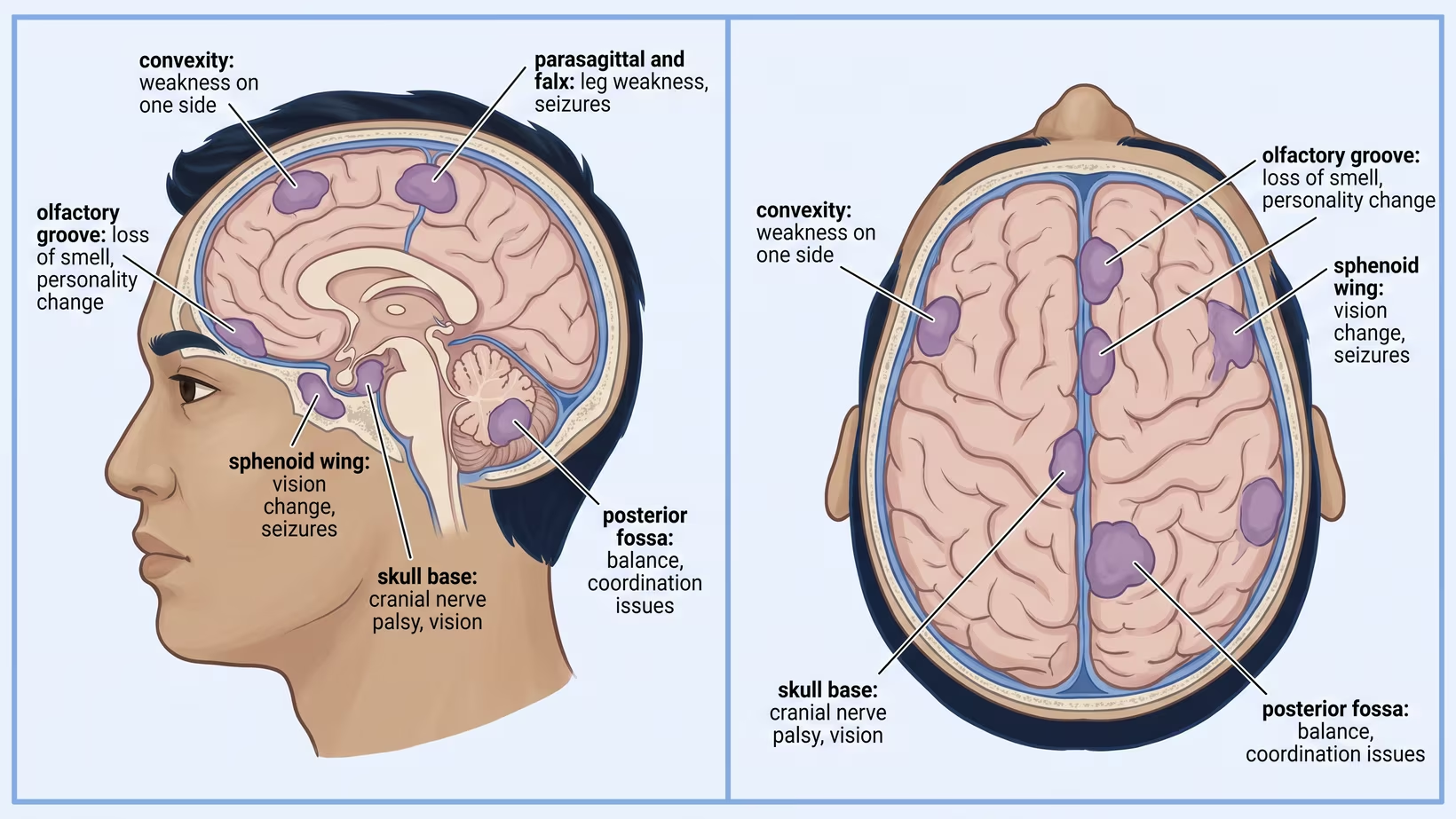
Location decides almost everything practical: which symptoms appear, how difficult an operation would be, and whether radiation is an option at all. Two tumors of identical size behave completely differently in different places.
The names that appear in reports describe position. A convexity meningioma sits over the outer surface of the brain. Parasagittal and falx tumors sit near the midline, beside the large vein that drains blood from the top of the brain. A sphenoid wing tumor sits behind and beside the eye. An olfactory groove tumor sits underneath the front of the brain, near the nerves for smell. Skull base and posterior fossa tumors sit deep, close to the cranial nerves, the brainstem and the balance pathways.
Swelling in the brain next to the tumor matters as much as the tumor. Seizures are much more likely when that swelling is present, and more likely when the tumor is not at the skull base.
In an older adult, gradual change is worth a scan whatever the cause, because slow pressure has more than one explanation. It may be gradual change in an older adult that turns out to be a chronic subdural hematoma rather than a tumor.
What happens after a meningioma diagnosis?
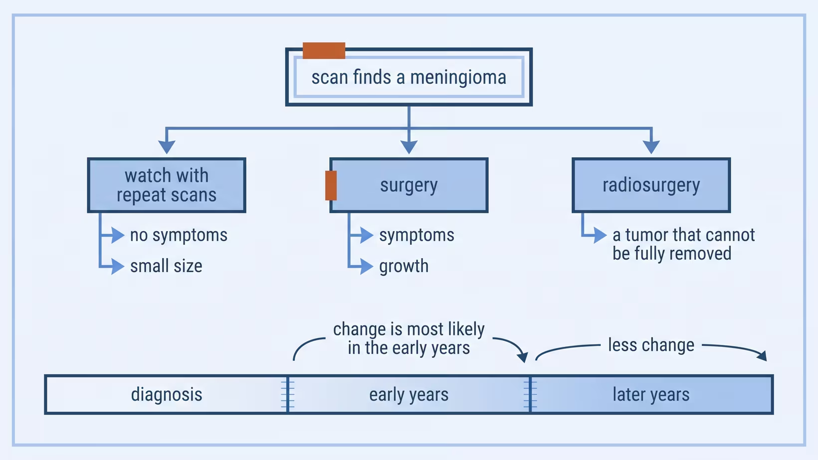
A meningioma is usually first identified on MRI, and very often that scan was ordered for another reason entirely. What follows is a review of the imaging in clinic, and then one of three paths: watching with repeat scans, surgery, or radiosurgery.
What decides between them is the combination of whether the tumor is causing anything, its size, exactly where it sits, whether there is swelling around it, whether it has grown since an earlier scan, and the patient’s age and general health. Removing the tumor completely, including the piece of covering it grew from, is often curative. When a tumor cannot be removed, or comes back and needs treatment, radiosurgery is used where the size and the nearby structures allow it, and fractionated radiotherapy where they do not. No drug is established as a standard treatment.
If watching is the plan, the neurosurgeon sets the interval between scans. The early years are when change is most likely, so the first few appointments are the ones not to skip.
Does a meningioma always need surgery?
No. Watching with repeat scans is a recognised plan rather than neglect, and it is used particularly when a tumor is causing no symptoms, when the patient is elderly, or both.
The reasoning is straightforward. Among incidental meningiomas that are monitored, only a small minority ever cause symptoms, and about a quarter eventually come to treatment. Operating on every tumor at the moment it is found means operating on many that never needed it.
Waiting is the wrong choice in other situations. A surgeon is unlikely to advise watching when the tumor is already causing symptoms, when comparison with an earlier scan shows it has grown, when there is swelling in the brain around it, when it is large, or when it sits somewhere that a small amount of further growth would threaten vision or another critical function.
Whichever way your own surgeon advises, that advice is built on your scan and your examination, which this page does not have.
Bring and ask
Questions to ask your doctor about a meningioma
Write these down before the consult, or screenshot them. A written list gets more useful answers in less time than trying to remember on the day.
- What grade is suspected, and how will that be confirmed
- Exactly where is it sitting, and what important structures are next to it
- Is there swelling in the brain around it
- Is the plan to watch, to operate, or to consider radiation, and why that one
- If we watch, when is the next scan, and what are we comparing it against
- What would make this urgent enough for me to call before that
- Who do I call at night or on a weekend
- Does anything need to change about driving, work, or being alone
Bring the actual images, every prior scan you can find, a list of your current medicines, and one person who will remember the conversation. For what else the first visit involves, see what to expect and bring to a first neurosurgery appointment.
What to ask about cost and PhilHealth coverage
There is no honest single figure to publish here, because the cost depends on the hospital, the room, the imaging and the plan, and any number printed on a page goes stale. What you can do is ask the right offices, in the right order, and get numbers that are actually yours.
Ask the hospital billing or social service office for a written estimate, keeping the workup and the operation separate. Ask what the estimate assumes about the room, and whether it includes the surgeon, the anaesthesiologist, implants, intensive care, and pathology. Ask what happens to the estimate if the stay runs longer than planned.
Ask the PhilHealth desk at the hospital what applies to this admission, and exactly which documents and contributions they need from you. Ask the surgeon’s secretary about professional fees and about what is paid before admission.
What moves the total most: the room, how much imaging is repeated, whether radiation is part of the plan, and how long the stay turns out to be.
The referral path for a meningioma in the Philippines
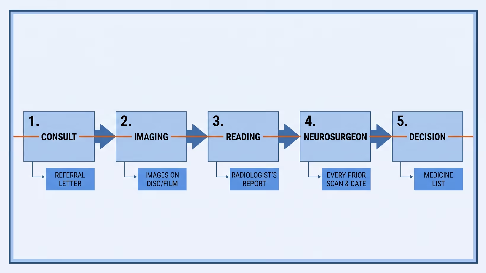
The usual route runs in one direction. The first doctor you see orders the scan, a radiologist reads it, and a neurosurgeon decides what happens next. A family physician, internist or neurologist can start that route from anywhere in the country.
The queue is real, and it is worth understanding rather than resenting. As reported in 2020, there were about 134 certified neurosurgeons in the Philippines, roughly one for every 800,000 people, trained in ten centres, with about three new neurosurgeons added each year. That is the reason to arrange the first consult when the scan is found rather than when symptoms change.
From a provincial or primary care setting, three things make the trip count. Ask your doctor for a referral letter that names the finding and the specific question being asked. Carry the images themselves, not only the report. Bring any older scan, however unrelated it seemed at the time.
What should a GP do with a meningioma found on a scan?
An incidental meningioma in a patient with no symptoms is not an emergency, and it does not need treatment started in primary care. The useful work is documentation, the right imaging, and a referral that arrives with enough information to be acted on.
Document a baseline neurological examination, including visual fields and gait, plus functional status and any seizure history. That baseline is what a change is later measured against, and nobody can reconstruct it afterwards.
The provisional diagnosis rests on MRI rather than CT alone. A report is most useful to the surgeon when it states the size, the exact location and the dural attachment, the relationship to the venous sinuses and cranial nerves, and whether there is swelling in the brain around the tumor. Note the date of every prior scan, since progression is judged by comparison.
Do not start an anticonvulsant in a patient who has never had a seizure. The available evidence does not support routine preventive use, and a meningioma on its own is not an indication.
What to tell the patient meanwhile: the grade cannot be read off the scan, most of these grow slowly, the plan is a neurosurgical opinion rather than an immediate operation, and these specific signs mean coming back sooner.
When to refer, and what to send with the patient
Two lists, kept apart, because they carry different urgency.
Refer the same day or the next:
- A first seizure
- A neurological deficit that has progressed over days to weeks
- Reduced alertness or new confusion
- New visual loss, or a visual field defect on examination
- Headache with vomiting, or headache worse on lying flat
Refer routinely:
- Incidental finding, no symptoms, normal examination
- Stable findings with old imaging available for comparison
- A patient who needs the plan explained more than they need an operation
Send with the patient:
- The images themselves, on disc or film, not only the radiologist’s report
- The date of every prior scan, so growth can actually be measured
- Your neurological findings, written down
- Seizure history, including single events years ago
- The full medicine list, naming any anticoagulant or antiplatelet
- Comorbidities that would affect anaesthesia
- A contact number for you that is answered
Frequently asked questions
Did something I did cause this?
In most people the cause of a meningioma is never identified. Stress, mobile phones and food have not been shown to cause it. Tell your doctor if you ever had radiation to the head or take a hormone medicine long term.
Can a meningioma be cured?
Complete removal of the tumor together with the covering it grew from is often curative. That is not a promise for any individual, and follow-up scans continue afterwards to make sure.
Will I need radiation?
Not everyone does. Radiosurgery or fractionated radiotherapy is used when a tumor cannot be removed completely, or when it returns and needs treatment.
Can medicine shrink a meningioma?
No drug is established as a standard treatment for meningioma. Medicines are used to control symptoms such as seizures, not to remove the tumor.
Is a meningioma hereditary?
For most patients no inherited cause is found. Tell your doctor if other relatives have had tumors of the brain or nerves, because a family pattern changes what a specialist looks for.
Should a healthy person get a scan to check?
No. Tumor type findings do turn up on brain MRI in people with no symptoms, more often with older age and with higher resolution scanners, but that is not a reason to screen healthy people.
Can a child have a meningioma?
Yes, though far less often than an adult. It is managed by a neurosurgeon, with the plan built on the location and what the tissue shows, and the specialist may also check for an inherited cause.
Can I still work or drive?
That depends on your symptoms, especially whether you have had a seizure, and it is a question for the doctor who knows your case. Ask specifically about driving, working at height, swimming alone, and night shifts.
See sources
- Goldbrunner R et al, for the European Association of Neuro-Oncology. EANO guideline on the diagnosis and management of meningiomas. Neuro-Oncology, 2021. PMID 34181733, doi:10.1093/neuonc/noab150
- Price M, Ostrom QT et al, Central Brain Tumor Registry of the United States. CBTRUS Statistical Report: Primary Brain and Other Central Nervous System Tumors Diagnosed in the United States in 2017-2021. Neuro-Oncology, 2024. PMID 39371035, doi:10.1093/neuonc/noae145
- Islim AI, Jenkinson MD et al. Incidental intracranial meningiomas: a systematic review and meta-analysis of prognostic factors and outcomes. Journal of Neuro-Oncology, 2019. PMID 30656531, doi:10.1007/s11060-019-03104-3
- Englot DJ et al. Seizures in supratentorial meningioma: a systematic review and meta-analysis. Journal of Neurosurgery, 2016. PMID 26636386, doi:10.3171/2015.4.JNS142742
- Morris Z, Al-Shahi Salman R et al. Incidental findings on brain magnetic resonance imaging: systematic review and meta-analysis. BMJ, 2009. PMID 19687093, doi:10.1136/bmj.b3016
- Louis DN, Perry A, Cree IA et al. The 2021 WHO Classification of Tumors of the Central Nervous System: a summary. Neuro-Oncology, 2021. PMID 34185076, doi:10.1093/neuonc/noab106
- Ferraris KP, Vesagas T, Seng K et al. The state of neurosurgical training and education in East Asia: analysis and strategy development for this frontier of the world. Neurosurgical Focus, 2020. PMID 32114563, doi:10.3171/2019.12.FOCUS19814
- National Institute for Health and Care Excellence. Suspected cancer: recognition and referral, section 1.9 Brain and central nervous system cancers. NICE guideline NG12, 2015. https://www.nice.org.uk/guidance/ng12
- Weill A, Nguyen P, Labidi M et al. Use of high dose cyproterone acetate and risk of intracranial meningioma in women: cohort study. BMJ, 2021. PMID 33536184, doi:10.1136/bmj.n37
- Vienne-Jumeau A, Tafani C, Ricard D. Environmental risk factors of primary brain tumors: A review. Revue Neurologique, 2019. PMID 31526552, doi:10.1016/j.neurol.2019.08.004